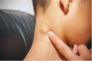
The majority of the time, swollen lymph nodes are a sign that your body is battling a virus or bacterial infection. Immune cells, viruses or bacteria, and fluid fill the nodes, increasing their size. Rarely, other, graver diseases can also result in swollen lymph nodes. Lymphadenopathy is fairly typical. Typically, it is not clinically significant in and of itself. However, it might indicate a serious underlying illness. For clinicians, the challenge is to avoid aggressive examination and biopsy of the majority of children while providing prompt, accurate for kids with serious underlying diseases. Lymphadenopathy is the term for swollen glands or swelling of the lymph nodes. The lymph glands are part of the immune system and help fight infections and other disease. They are enlarged when the body is fighting infection or other diseases. In children its to be able to feel some lymph nodes small, movable lumps under the skin. But if the nodes get bigger than usual, your child may have an infection or other problem. In most cases, lymphadenitis clears up quickly with proper treatment but it may take more time for lymph nodes swelling to go away. Be sure to let your healthcare provider know if your lymphadenitis symptoms come back. The most common treatment for swollen lymph nodes caused by a bacterial infection is antibiotics.
This is an Open Access article, distributed under the terms of the Creative Commons Attribution 4.0 International License (http://creativecommons.org/licenses/by/4.0/), which permits unrestricted use, distribution and reproduction in any medium or format, provided the original work is properly cited.
1) Swollen lymph nodes are a typical sign in clinical settings. The reticuloendothelial system's lymph nodes, which drain fluid from the majority of body tissues, are a target for a variety of antigens.
2) A wide range of conditions, from more serious infections and malignancies to self-limited viral illnesses, can result in lymphadenopathy.
3) Children have physiological lymphadenopathy, unlike adults.
4) Knowledge of the disease's normal anatomic drainage, physiological changes, and epidemiological aspects aids in accurate diagnosis, wise test selection, and avoidance of hasty treatment.
2. Epidemiology
Acute, subacute, or chronic lymphadenopathy are all possible. Because they resolve on their own, many causes of acute and subacute lymphadenopathy may go unnoticed. Chronic lymphadenopathy is worrying and frequently requires a precise diagnosis. It is challenging to pinpoint the exact cause of lymphadenopathy. Non-tuberculous mycobacteria are the most frequent cause of chronic lymphadenopathy in developed nations with low Mycobacterium tuberculosis (MTB) prevalence, whereas tuberculosis is the most frequent cause of chronic lymphadenopathy in India. The most frequent cause is reactive lymphadenitis.
[1]
Reuss AM, Wiese-Posselt M, Weimann B, et al. Incidence rate of nontuberculous mycobacterial disease in immune ocompetent children a prospective nationwide surveillance study in Germany. PediatrInfect Dis J. 2009; 28(7): 642-4.
Northeastern India has a higher prevalence of endemic fungal infections, such as sporotrichosis, cryptococcosis, and blastomycosis. West Bengal, Uttar Pradesh, and some of the plains of the Gange are endemic to histoplasmosis. The actual incidence of these infections is unknown because they are non-reportable illnesses.
[4]
Chakrabarti A, Slavin MA. Endemic fungal infections in the Asia-Pacific region. Med Mycol. 2011; 49(4): 337-44.
If a lymph node's diameter exceeds 1 cm, it is deemed severely enlarged. Superior helix nodules larger than 0.5 cm and inguinal nodules larger than 1.5 cm are exceptions to this definition. Any size of the popliteal and supraclavicular tubercles that can be felt is always considered significant.
[5]
Bruce shiraizu MG. Evaluation of lymphadenopathy in children. CurrOpinpediatr. 1994; 6(1): 68-76.
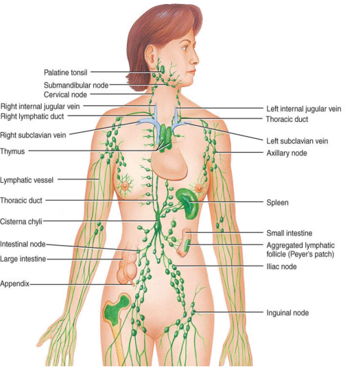
Lymphadenopathy can be physiological or secondary to childhood antigenic exposures that were frequent and varied, infections from bacteria or viruses, cancer, drug-induced disorders, storage disorders, etc. Finding the cause of focal lymphadenitis can be made easier with knowledge of the lymph node groups' draining patterns. The cervical lymph node group drains the areas of the head, neck, and throat. Younger children frequently experience pharyngitis, otitis media, and scalp infections that result in enlarged neck lymph nodes. The axillary lymph node group receives drainage from the upper extremities, upper abdomen, and superficial and lateral areas of the chest wall. The groin lymph node group receives drainage from the lower extremities, the genitourinary system, and the perineum. Enlargement of the supraclavicular lymph nodes is always cause for concern and should prompt a thorough examination for malignancies, fungi, and tuberculosis. The age of the child, underlying immunological status (immunocompetent or immunocompromised), and location (endemic) are all variables that may help identify the etiology of lymphadenopathy. Environmental exposure (Cryptococcus eucalyptus trees, histoplasma excavation and activity in caves and logs, meliidosis, soil-contaminated wounds), travel to endemic areas, consumption of unpasteurized milk (brucellosis, Mycobacterium bovis), diseases like tuberculosis, shrub typhoid, and endemic fungi are just a few examples. ingesting raw meat (Toxoplasma) and coming into contact with animals and birds (Bartonella Histoplasma, Cryptococcus, and Toxoplasma). If they exist, these are significant indicators, but it can be challenging to spot them unless your doctor specifically asks for them.
[6]
Mohseni S, Khorgami Z, Alinejad S, et al. Peripheral Lymphadenopathy: Approach and Diagnostic Tools. Iran J Med Sci. 2014; 39(2): 158-70.
An organized description of the lymph nodes can help to focus the diagnosis and enable targeted testing when examining kids with lymphadenopathy. Size, number, tenderness, temperature, hardness, area, mat, exit nasal cavity, associated lymphangitis, motility, duration of symptoms (acute, subacute 1-3 weeks, chronic - >4 weeks), localization (local vs. generalized), etc. Fixation is a crucial factor to take into account because these results point to causality. Performance status, anemia, cutaneous hemorrhages (suggestive of bone marrow infiltrative processes like leukemia), and examination of the conjunctiva (gloal glial syndrome) are among the additional findings assessed as part of the general and systemic evaluation. Cat scratch disease, herpes simplex virus (HSV), tularemia, adenovirus, pyogenic bacterial infection, ear/throat, erythema nodosum, chest findings (abnormal breath sounds, pleura), pleural effusion, consolidation, fibrosis), and hepatosplenomegaly (occurring in many systemic infections).
[7]
Stutchfield CJ, Tyrrell J. Evaluation of lymphadenopathy in children. Paediatr Child Health. 2012; 22(3): 98-102.
The number of lymph nodes involved, the location of the lymphadenopathy, and the length of the symptoms all have an impact on the etiology. "Based on the collection of associated lymph nodes, Table 1 lists the most frequent causes; however, the list is not all-inclusive." 8,9 Papules, scabs, and ulcers are visible in cases of cat scratch disease, typhoid diphtheria, anthrax, pasteurellamultocida, nocardiosis, and tick fever.
[10]
Kliegman R, Lye PS, Bordini BJ, et al. Nelson pediatric symptom- based diagnosis. 2018. Available from:
Table 2. Antibodies to Epstein-Barr virus in EBV infection.
Infection
VCAIgM
VCAIgG
Early antigen
EBNA
Acute infection
+
+
+/-
-
Recent infection
+/-
+
+/-
+/-
Past infection
-
+
+/-
+
5.1. Diagnosis
A thorough history and testing are all that are required to diagnose acute focal lymphadenitis. Blood counts and inflammatory markers may show elevated ESR and C-reactive protein (CRP), left-shifted values, but they don't really help with clinical evaluation. A swinging knot can imply sensibility and culture. We can cultivate microorganisms that allow us to choose antibiotics with the right level of sensitivity. Antibiotic use in the past frequently makes the culture sterile. A thorough examination of the lymph nodes in the supraclavicular region is always necessary. Acute focal lymphadenitis is best treated with no or little intervention, whereas subacute, chronic, and generalized lymphadenopathy require specialized diagnostic testing to identify the underlying cause.
5.2. Hematology
ESR, CRP, LDH, and complete blood count all aid in excluding malignancy. Even though they are non-specific, elevated inflammatory markers may point to an infectious cause. Blasts, cytopenia, and an elevated LDH level point to a malignant etiology. DNA testing and serology: The most frequent cause of acute pharyngitis and tender neck lymphadenopathy in group A children requiring antibiotic therapy is streptococcal pharyngitis. GAS pharyngitis can be identified using a throat swab culture or a rapid streptococcal antigen test (RSAT).
[11]
Gerber MA, Shulman ST. Rapid Diagnosis of Pharyngitis Caused by Group A Streptococci. ClinMicrobiol Rev. 2004; 17(3): 571-80.
Pharyngitis caused by the Epstein-Barr virus and its associated lymphadenopathy are identical to those caused by GAS. The use of serological testing can help (Table 2) distinguish between primary EBV infections and earlier infections.
[12]
Kimberlin DW, Brady MT, Jackson MA, et al. Author: Committee on Infectious Diseases, Am AcadPediatr. 2013; 57(8): 1145-54.
[12]
The medical history, conventional laboratory results, immunofluorescent antibody testing (IFA), and PCR are used to make the diagnosis of cat scratch disease. Regular cultivation techniques rarely result in success.
[13]
Chaudhry R, Kokkayil P. Ghosh A, et al. Bartonellahenselae infection in diverse clinical conditions in a tertiary care hospital in north India. Indian J Med Res. 2018; 147(2): 189-94.
Children are more commonly affected by glomers than adults, who are more commonly affected by bloodstream infections. The most accurate way to diagnose smallpox is through a culture of a blood or tissue sample.
However, the turnaround time he required was more than a week, and only knowledgeable inspectors could make the identification.
[15]
Lumbiganon P, Chotechuangnirun N. Kosalaraksa P, et al. Localized Melioidosis in Children in Thailand: Treatment and Long-term Outcome. J Trop Pediatr. 2011; 57(3): 185-91.
Inglis TJJ, Merritt A, Chidlow G, et al. Comparison of Diagnostic Laboratory Methods for Identification of BurkholderiapseudomalleiJClinMicrobiol. 2005; 43(5): 2201-6.
Urinary Histoplasma Antigen Test and Serum/CSF Cryptococcal Antigen Test (CrAg) can be used for a quick diagnosis with excellent sensitivity and specificity.
5.3. Imaging Tests
The diagnosis of associated hilar/mediastinaladenopathy is assisted by chest x-rays and CT scans. Mediastinal/hilar lymph nodes are linked to tuberculosis, histoplasmosis, blastocycosis, and coccidioidomycosis.
Based on the characteristics of the nodes, ultrasound can distinguish infected from malignant nodes and distinguish lymph nodes from other swellings. Identification of necrotic intra-abdominal nodules and hepatosplenic lesions linked to tuberculosis, melioidosis, and brucellosis is aided by abdominal ultrasonography.
[17]
Tschammler A, Ott G, Schang T, et al. Lymphadenopathy: differentiation of benign from malignant disease-color Doppler US assessment of intranodalangioarchitecture. Radiology. 1998; 208(1): 117-23.
5.4. Fine Needle Aspiration Cytology (FNAC) and Biopsy
Most cases of chronic lymphadenopathy in kids and teenagers are benign. Due to its minimal invasiveness, cost-effectiveness, ready availability, lack of general anesthesia, and low associated morbidity, FNAC is the preferred method for histopathological diagnosis. In between 10 and 20 percent of FNAC samples, chronic sinus formation indicative of tuberculosis and NTM disease may be non-diagnostic. Because an open biopsy, unlike FNAC, can provide enough specimen for culture, PCR, and histopathology, doctors shouldn't be afraid to perform one even if FNAC is inconclusive.
[8]
Twist CJ, Link MP. Assessment of lymphadenopathy in children. PediatrClin North Am. 2002; 49(5): 1009-25.
acute viral gonadoinflammatory syndrome Reassuring caregivers and educating them on the warning signs for prompt reassessment is the best course of action if the history and physical examination indicate benign viral lymphadenopathy. Unless they have severe liver or respiratory disease, children rarely require specific antiviral therapy.
[9]
Gosche JR, Vick L. Acute, subacute, and chronic cervical lymphadenitis in children. SeminPediatr Surg. 2006; 15(2): 99-106.
A bacterial infection is frequently to blame for a sensitive neck lump that is enlarged to one side. In children ages 1-4, Staphylococcus aureus and GAS account for the majority of cases. In older kids, oral anaerobes are linked to periodontitis. These widespread pathogens should be the focus of initial antibiotic treatment. Amoxicillin and clavulanate are frequently used because penicillinase-producing staphylococci are so prevalent. Typically, a course of treatment lasts 10 days. In India, community-acquired MRSA is on the rise. If there is a high prevalence of clindamycin-resistant MRSA, adding clindamycin is the best empirical treatment. MANY doctors prefer azithromycin because it is simple to administer once daily, but the dosage needs to be 12 mg/day. kg/day. Macrolides should not be used as the first-line treatment for bacterial lymphadenitis due to the persistent rise in GAS resistance to macrolide gases in India. If there is no reaction or persistent lymphadenopathy, further testing is necessary. Adenitis due to acute bacteria. A bacterial infection is frequently to blame for a sensitive neck lump that is enlarged to one side. In children ages 1-4, Staphylococcus aureus and GAS account for the majority of cases. In older kids, oral anaerobes are linked to periodontitis. These widespread pathogens should be the focus of initial antibiotic treatment. Amoxicillin and clavulanate are frequently used because penicillinase-producing staphylococci are so prevalent. Typically, a course of treatment lasts 10 days. In India, community-acquired MRSA is on the rise. If there is a high prevalence of clindamycin-resistant MRSA, adding clindamycin is the best empirical treatment.
[18]
Alvarez-Uria G, Reddy R. Prevalence and Antibiotic Susceptibility of Community-Associated Methicillin-Resistant Staphylococcus aureus in a Rural Area of India: Is MRSA Replacing Methicillin- Susceptible Staphylococcus aureus in the Community? ISRN Dermatol. 2012; 2012: 1-5.
Many doctors prefer azithromycin because it is simple to administer once daily, but the dosage must be 12 mg/day, kg/day. Macrolides shouldn't be the first-line treatment for bacterial lymphadenitis because GAS resistance to them is increasing year by year in India.
[19]
Brahmadathan KN, Anitha P, Gladstone P, et al. Increasing erythromycin resistance among group A streptococci causing tonsillitis in a tertiary care hospital in southern India. ClinMicrobiol Infect. 2005; 11(4): 335-7.
Further assessment is necessary if the treatment is unsuccessful or the lymphadenopathy persists.
6.3. Subacute and Chronic Lymphadenitis
It can be brought on by a variety of non-infectious conditions, including tuberculosis, non-tuberculous mycobacterial disease, cat scratch disease, meliidosis, toxoplasmosis, fungal infections, and parasitic infections. Epidemiologically or clinically, it is impossible to distinguish between NTM lymphadenitis and tuberculous lymphadenitis. It is covered elsewhere how to treat tuberculous lymphadenitis. Surgical intervention, either with or without antibiotics, is the mainstay of treatment for NTM lymphadenitis. The most frequent NTM associated with lymphadenitis in India are M. avium complex (MAC), M. fortuitum, M. chelonae, and M. kansasii. The isolation rate of NTM in India is 2.6-3.9%.
[20]
Kishore Reddy VC, Prasad CE, Aparna S, et al. A study of mycobacterial species causing Lymphadenitis. Southeast Asian J Trop Med Public Health. 2008; 39(1): 130-5.
[20]
The preferred course of treatment for NTM lymphadenitis is resection, either with or without chemotherapy. Antimicrobial susceptibility is needed in order to direct treatment because NTM is resistant to common tuberculosis medications. A combination of rifampicin, ethambutol, and clarithromycin/azithromycin is recommended for the treatment of MAC-related recurrent disease and difficult-to-remove nodules. Three to six months are needed for treatment. Treatment for M. fortuitum lymphadenitis should last at least 4 months and involve at least 2 susceptible pathogens. shape. A 4-month course of chelonae combination therapy involving tobramycin, meropenem/linezolid, and clarithromycin is advised. M. kanasasi lymphadenitis may be treated with a daily regimen of rifampicin, isoniazid, and ethambutol for a period of six months.
[12]
Kimberlin DW, Brady MT, Jackson MA, et al. Author: Committee on Infectious Diseases, Am AcadPediatr. 2013; 57(8): 1145-54.
[21]
Griffith DE, Aksamit T, Brown-Elliott BA, et al. An Official ATS/ IDSA Statement: Diagnosis, Treatment, and Prevention of Nontuberculous Mycobacterial Diseases. Am J RespirCrit Care Med. 2007: 175(4): 367-416.
Cat scratch disease (localized lymphadenitis) typically resolves on its own within two to four months. However, the largest study evaluating 268 patients with Bartonella infection showed that the mean illness duration among treated individuals was 2.8 weeks compared to 14.5 weeks in the nontreated group. Antimicrobial therapy for the treatment of localized lymphadenitis is controversial.
[23]
Margileth AM. Antibiotic therapy for cat-scratch disease: clinical study of therapeutic outcome in 268 patients and a review of the literature. Pediatr Infect Dis J. 1992; 11(6): 474-8.
[23]
Azithromycin is the recommended first-line antibiotic, followed by doxycycline for 7–10 days. Clarithromycin, ciprofloxacin, trimethoprim-sulfamethoxazole (TMP SMX), and rifampicin are additional substitute antibiotics.
Children with localized melioidosis may benefit from receiving IV ceftazidime for 14 days, followed by 3-6 months of oral TMP-SMX alone or in combination with Doxycycline (age >8 years). The likelihood of recurrence increases with shorter antibiotic courses.
[24]
Mukhopadhyay C, Eshwara V, Kini P, et al. Pediatric Melioidosis in Southern India. Indian Pediatr. 2015; 52(8): 711-2.
[24]
The majority of toxoplasma infections acquired postnatallydon't need to be treated. However, pyrimethamine sulfadiazine should be used to treat immunosuppressed children and immunocompetent children with severe primary toxoplasmosis. There is no pyrimethamine sulfadiazine in India. In several case reports, patients with HIV were successfully treated with TMP-SMX for central nervous system toxoplasmosis. He will require antibiotics for 4-6 weeks after the symptoms go away.
[25]
Patil V, Rajmane V. Raje V, et al. Successful treatment of cerebral toxoplasmosis with cotrimoxazole. Indian J Sex Transm Dis AIDS. 2011; 32(1): 44-6.
[25]
The severity of the disease and the child's immunological state both influence how the disease is treated. Immunocompetent patients with non-meningeal disease and pulmonary involvement can be treated with fluconazole plus flucytosine for a period of three to six months. Itraconazole is a potential substitute. An initial induction phase with intravenous administration is necessary for the treatment of immunocompromised children. Flucytosine and amphotericin for two weeks came next.
Itraconazole 5 mg/kg/dose twice daily (maximum 200 mg/dose) for 2-4 weeks or until all lesions disappear is the preferred treatment for localized and uncomplicated sporotrixis.
[26]
Mahajan VK. Sporotrichosis: An Overview and Therapeutic Options. Dermatol Res Pract. 2014: 2014: 1-13.
[26]
The illness histoplasmosis is self-limiting. Children with immune compromise who have mild to moderate disease do not need to be treated because they have an acute lung disease. Itraconazole can be administered for 6 to 12 weeks if the patient's condition does not improve after 4 weeks. In disseminated cases and in patients with underlying immunodeficiency, an initial course of treatment with intravenous amphotericin followed by oral itraconazole is necessary.
[27]
Gopalakrishnan R, Nambi PS, Ramasubramanian V, et al. Histoplasmosis in India: Truly Uncommon or Uncommonly Recognised? J Assoc Physicians India. 2012; 60: 25-8.
[27]
7. Result
An abnormal physical finding in another system is frequently present with generalized lymphadenopathy, which is brought on by a systemic illness. Local lymphadenopathy: Usually caused by an infection of the lymph node or the area it drains from. When a nonbacterial infectious agent causes lymphadenopathy, it is characterized by an unusual anatomic site, a protracted course, sinus drainage, the absence of a prior pyogenic infection, and unusual antecedent disease.
8. Discussion
Parents frequently take their kids to the pediatrician's office when they discover a lump in their child's neck, armpit, or groin. They worry and inquire as to whether it is cancer. Even though the doctor has told the parents the child is not seriously ill, parents frequently seek a second or third opinion weeks or months after the lump has not subsided. They ask for a specific diagnosis and the anticipated cure date for the illness. It is our duty as first responders to examine this lump, describe it to the parents, and administer the proper care. When are these knots actually important, and when should you be concerned?The current literature review and our clinical expertise served as the foundation for this article. This article gives an overview of childhood lymphadenopathy and evaluation tips for this typical childhood ailment.
9. Conclusion
Although lymphadenopathy is a common condition, there are numerous possible causes for it. However, thorough understanding of physiologic variations, epidemiological factors, and a sound medical history and testing can assist clinicians in reducing the number of possible diagnoses. Ideally, medical professionals shouldn't be reluctant to examine nodules histopathologically.
Abbreviations
NTM: Non-Tuberculos Mycobacteria
HIV: human Immunodeficiency Virus
EBV: Epstein-Barr Virus
CMV: Cytomegalo Virus
HSV: Herpes Simplex Virus
Acknowledgments
I would like to express my special thank of gratitude to the management of AmepurvaForum, Nirant institute of Pharamacy, Boramani, Solapur.
I would also like to thankteachingstaffandparents and friends who helped me a lot in finalizing this paper on lymphadenopathy in child carewithin limited time frame.
Conflicts of Interest
The authors declare noconflicts of interest.
References
[1]
Reuss AM, Wiese-Posselt M, Weimann B, et al. Incidence rate of nontuberculous mycobacterial disease in immune ocompetent children a prospective nationwide surveillance study in Germany. PediatrInfect Dis J. 2009; 28(7): 642-4.
Kimberlin DW, Brady MT, Jackson MA, et al. Author: Committee on Infectious Diseases, Am AcadPediatr. 2013; 57(8): 1145-54.
[13]
Chaudhry R, Kokkayil P. Ghosh A, et al. Bartonellahenselae infection in diverse clinical conditions in a tertiary care hospital in north India. Indian J Med Res. 2018; 147(2): 189-94.
Lumbiganon P, Chotechuangnirun N. Kosalaraksa P, et al. Localized Melioidosis in Children in Thailand: Treatment and Long-term Outcome. J Trop Pediatr. 2011; 57(3): 185-91.
Inglis TJJ, Merritt A, Chidlow G, et al. Comparison of Diagnostic Laboratory Methods for Identification of BurkholderiapseudomalleiJClinMicrobiol. 2005; 43(5): 2201-6.
Tschammler A, Ott G, Schang T, et al. Lymphadenopathy: differentiation of benign from malignant disease-color Doppler US assessment of intranodalangioarchitecture. Radiology. 1998; 208(1): 117-23.
Alvarez-Uria G, Reddy R. Prevalence and Antibiotic Susceptibility of Community-Associated Methicillin-Resistant Staphylococcus aureus in a Rural Area of India: Is MRSA Replacing Methicillin- Susceptible Staphylococcus aureus in the Community? ISRN Dermatol. 2012; 2012: 1-5.
Brahmadathan KN, Anitha P, Gladstone P, et al. Increasing erythromycin resistance among group A streptococci causing tonsillitis in a tertiary care hospital in southern India. ClinMicrobiol Infect. 2005; 11(4): 335-7.
Kishore Reddy VC, Prasad CE, Aparna S, et al. A study of mycobacterial species causing Lymphadenitis. Southeast Asian J Trop Med Public Health. 2008; 39(1): 130-5.
[21]
Griffith DE, Aksamit T, Brown-Elliott BA, et al. An Official ATS/ IDSA Statement: Diagnosis, Treatment, and Prevention of Nontuberculous Mycobacterial Diseases. Am J RespirCrit Care Med. 2007: 175(4): 367-416.
Margileth AM. Antibiotic therapy for cat-scratch disease: clinical study of therapeutic outcome in 268 patients and a review of the literature. Pediatr Infect Dis J. 1992; 11(6): 474-8.
[24]
Mukhopadhyay C, Eshwara V, Kini P, et al. Pediatric Melioidosis in Southern India. Indian Pediatr. 2015; 52(8): 711-2.
[25]
Patil V, Rajmane V. Raje V, et al. Successful treatment of cerebral toxoplasmosis with cotrimoxazole. Indian J Sex Transm Dis AIDS. 2011; 32(1): 44-6.
[26]
Mahajan VK. Sporotrichosis: An Overview and Therapeutic Options. Dermatol Res Pract. 2014: 2014: 1-13.
[27]
Gopalakrishnan R, Nambi PS, Ramasubramanian V, et al. Histoplasmosis in India: Truly Uncommon or Uncommonly Recognised? J Assoc Physicians India. 2012; 60: 25-8.
Kanna, L. S., Kulkarni, R. R., Bhanap, S. S. (2024). Determination of the Lymphadenopathy in Child Care. American Journal of Health Research, 12(2), 37-43. https://doi.org/10.11648/j.ajhr.20241202.13
Kanna, L. S.; Kulkarni, R. R.; Bhanap, S. S. Determination of the Lymphadenopathy in Child Care. Am. J. Health Res.2024, 12(2), 37-43. doi: 10.11648/j.ajhr.20241202.13
Kanna LS, Kulkarni RR, Bhanap SS. Determination of the Lymphadenopathy in Child Care. Am J Health Res. 2024;12(2):37-43. doi: 10.11648/j.ajhr.20241202.13
@article{10.11648/j.ajhr.20241202.13,
  author = {Laxmi Sidram Kanna and Rajashree Ramakant Kulkarni and Sakshi Swapnil Bhanap},
  title = {Determination of the Lymphadenopathy in Child Care
},
  journal = {American Journal of Health Research},
  volume = {12},
  number = {2},
  pages = {37-43},
  doi = {10.11648/j.ajhr.20241202.13},
  url = {https://doi.org/10.11648/j.ajhr.20241202.13},
  eprint = {https://article.sciencepublishinggroup.com/pdf/10.11648.j.ajhr.20241202.13},
  abstract = {The majority of the time, swollen lymph nodes are a sign that your body is battling a virus or bacterial infection. Immune cells, viruses or bacteria, and fluid fill the nodes, increasing their size. Rarely, other, graver diseases can also result in swollen lymph nodes. Lymphadenopathy is fairly typical. Typically, it is not clinically significant in and of itself. However, it might indicate a serious underlying illness. For clinicians, the challenge is to avoid aggressive examination and biopsy of the majority of children while providing prompt, accurate for kids with serious underlying diseases. Lymphadenopathy is the term for swollen glands or swelling of the lymph nodes. The lymph glands are part of the immune system and help fight infections and other disease. They are enlarged when the body is fighting infection or other diseases. In children its to be able to feel some lymph nodes small, movable lumps under the skin. But if the nodes get bigger than usual, your child may have an infection or other problem. In most cases, lymphadenitis clears up quickly with proper treatment but it may take more time for lymph nodes swelling to go away. Be sure to let your healthcare provider know if your lymphadenitis symptoms come back. The most common treatment for swollen lymph nodes caused by a bacterial infection is antibiotics.
},
 year = {2024}
}
TY  - JOUR
T1  - Determination of the Lymphadenopathy in Child Care

AU  - Laxmi Sidram Kanna
AU  - Rajashree Ramakant Kulkarni
AU  - Sakshi Swapnil Bhanap
Y1  - 2024/04/11
PY  - 2024
N1  - https://doi.org/10.11648/j.ajhr.20241202.13
DO  - 10.11648/j.ajhr.20241202.13
T2  - American Journal of Health Research
JF  - American Journal of Health Research
JO  - American Journal of Health Research
SP  - 37
EP  - 43
PB  - Science Publishing Group
SN  - 2330-8796
UR  - https://doi.org/10.11648/j.ajhr.20241202.13
AB  - The majority of the time, swollen lymph nodes are a sign that your body is battling a virus or bacterial infection. Immune cells, viruses or bacteria, and fluid fill the nodes, increasing their size. Rarely, other, graver diseases can also result in swollen lymph nodes. Lymphadenopathy is fairly typical. Typically, it is not clinically significant in and of itself. However, it might indicate a serious underlying illness. For clinicians, the challenge is to avoid aggressive examination and biopsy of the majority of children while providing prompt, accurate for kids with serious underlying diseases. Lymphadenopathy is the term for swollen glands or swelling of the lymph nodes. The lymph glands are part of the immune system and help fight infections and other disease. They are enlarged when the body is fighting infection or other diseases. In children its to be able to feel some lymph nodes small, movable lumps under the skin. But if the nodes get bigger than usual, your child may have an infection or other problem. In most cases, lymphadenitis clears up quickly with proper treatment but it may take more time for lymph nodes swelling to go away. Be sure to let your healthcare provider know if your lymphadenitis symptoms come back. The most common treatment for swollen lymph nodes caused by a bacterial infection is antibiotics.

VL  - 12
IS  - 2
ER  -