Abstract
Introduction: The Anterior Lateral thigh flap (ALT) free flap can be considered one of the reconstructive tools in lower leg and foot reconstruction. It has a long and reliable pedicle, and dissection is comparatively easy, which makes it extremely suitable as a free flap. This study aimed to evaluate the versatility of the flap in covering lower leg and foot defects. Methods: This was a prospective observational study conducted in the Department of Plastic Surgery, Rangpur Medical College Hospital, Rangpur, Bangladesh, from January 2023 to December 2023. In our study, we included 16 patients with defects in the lower leg and foot who received an ALT-free flap as a reconstructive treatment. We analyzed the preoperative, intraoperative, and postoperative variables of our respondents. Results: In this study, 16 patients (12 males and 4 females) with an average of 34.25 years old were included. Among them 13 were traumatic cases, 2 electrical burn wounds, and one oncologic case. Survival rates of the free flap were 87.5% (14) with two failure cases. There were no complications at the donor site. In this study, 75% (12) cases showed excellent outcomes where there were no signs, or symptoms of venous congestion or arterial insufficiency, no infection or dehiscence, and the donor site was primarily closed with good flap adhesion. Conclusion: This study shows that the ALT-free flap is a versatile flap that allows it to cover the defect successfully and ALT adaptation was satisfactory in terms of varied location and size.
Keywords
Anterolateral Thigh Flap, Split Thickness Skin Graft, Versatility, Foot Reconstruction
1. Introduction
The Anterior Lateral thigh flap
(ALT) free flap has become popular in Asian countries, where it has replaced the radial forearm flap to cover a variety of defects. Since its development in 1984 by Song et al., it has gained increasing appreciation irrespective of defects in lower limb reconstruction
[1] | Song Yg, Chen Gz, Song Yl. The Free Thigh Flap: A New Free Flap Based On The Septocutaneous Artery. Br J Plast Surg 1984; 37: 149–159. |
[1]
.
This flap is based on septocutaneous branches of the descending branches of the lateral circumflex femoral artery. It is noted that the vascular anatomy was variable and the majority of skin vessels were septocutaneous compared to musculocutaneous. This flap is a so-called workhorse flap for the reconstruction of small or large defects and extensively reported in the various literature
[2] | Mardini S, Lin Ch, Wei Fc. In: Blondeel Pn, Morris Sf, Hallock Gg, Et Al, Eds. Perforator Flaps: Anatomy, Technique & Clinical Applications. St. Louis, Mo: Quality Medical Publishing; 2006: 617–633. |
[2]
.
Significant advantages characterize the ALT flap as versatility, easy dissection, adequate size to cover large wounds, and excellent perfusion. When the vessels near the wound are compromised, a long pedicle can be required to reach a distant blood source. ALT pedicle ranges from 7 cm to 9cm.
The ALT flap is useful for reconstruction in a variety of defects, including the head and neck, trunk, and upper extremities
[3] | Nosrati N, Chao Ah, Chang Dw, Yu P. Lower Extremity Reconstruction With The Anterolateral Thigh Flap. Journal Of Reconstructive Microsurgery. 2012 May; 28(04): 227-34. |
[4] | Lee N, Roh S, Yang K, Kim J. Reconstruction Of Hand And Forearm After Sarcoma Resection Using Anterolateral Thigh Free Flap. J Plast Reconstr Aesthet Surg 2009; 62(12): E584–E586. |
[3, 4]
. It has been proven to be useful in the healing of infected wounds due to its rich vascular supply
[5] | Gravvanis A, Tsoutsos D, Karakitsos D, Iconomou T, Papadopoulos O. Blood Perfusion Of The Free Anterolateral Thigh Perforator Flap: Its Beneficial Effect In The Reconstruction Of Infected Wounds In The Lower Extremity. World J Surg 2007; 31(1): 11–18. |
[5]
. Furthermore, the ALT flap can be combined with a vastus lateralis (VL) flap to add bulk or a tensor fascia lata flap for tendon repair
[6] | Kuo Yr, Kuo Mh, Chou Wc, Liu Yt, Lutz Bs, Jeng Sf. One-Stage Reconstruction Of Soft Tissue And Achilles Tendon Defects Using A Composite Free Anterolateral Thigh Flap With Vascularized Fascia Lata: Clinical Experience And Functional Assessment. Ann Plast Surg 2003; 50(2): 149–155. |
[6]
. The characteristics of the ALT flap—its versatility in thickness, potential size, and flexibility—also make it ideal for lower extremity reconstruction
[7] | Heller L, Kronowitz Sj. Lower Extremity Reconstruction. J Surg Oncol 2006; 94(6): 479–489. |
[7]
. Most studies of outcomes of lower extremity reconstruction with the ALT flap have focused on its use as a free flap for relatively distal defects
[8] | Park Je, Rodriguez Ed, Bluebond-Langer R, Et Al. The Anterolateral Thigh Flap Is Highly Effective For Reconstruction Of Complex Lower Extremity Trauma. J Trauma 2007; 62(1): 162–165. |
[9] | Demirtas Y, Neimetzade T, Kelahmetoglu O, Guneren E. Comparison Of Free Muscle And Perforator Skin Flaps For Soft Tissue Reconstruction Of The Foot And Ankle. Foot Ankle Int 2010; 31(1): 53–58.. |
[10] | Hong Jp, Kim Ek. Sole Reconstruction Using Anterolateral Thigh Perforator Free Flaps. Plast Reconstr Surg 2007; 119(1): 186–193. |
[8-10]
. The ALT flap has also been used as a pedicled flap for repair of more proximal lower extremity defects, though studies of this use often included patients with defects of the abdominal wall, perineum, and back
[11] | Ng Rw, Chan Jy, Mok V, Li Gk. Clinical Use Of A Pedicled Anterolateral Thigh Flap. J Plast Reconstr Aesthet Surg 2008; 61(2): 158– 164. |
[12] | Friji Mt, Suri Mp, Shankhdhar Vk, Ahmad Qg, Yadav Ps. Pedicled Anterolateral Thigh Flap: A Versatile Flap For Difficult Regional Soft Tissue Reconstruction. Ann Plast Surg 2010; 64(4): 458–461. |
[13] | Gravvanis Ai, Tsoutsos Da, Karakitsos D, Et Al. Application Of The Pedicled Anterolateral Thigh Flap To Defects From The Pelvis To The Knee. Microsurgery 2006; 26(6): 432–438. |
[11-13]
.
This study aimed to revise our experience with the use of ALT-free flap and assess its versatility in a group of patients with coverage defects of different origins, sizes, and locations in lower limbs
[14] | Wei Fc, Jain V, Celik N, Chen Hc, Chuang Dc, Lin Ch. Have We Found An Ideal Soft-Tissue Flap? An Experience With 672 Anterolateral Thigh Flaps. Plastic And Reconstructive Surgery. 2002 Jun 1; 109(7): 2219-26. |
[15] | Biswas D, Kalam Ma, Ahmed T, Khan Mrk. Results Of Microvascular Free Flap For Reconstruction Of Soft Tissue Defects: Our Early Experience. Bdjps 2012; 3(2): 33-37. |
[14, 15]
.
2. Methodology & Materials
This was a prospective observational study conducted in the Department of Plastic Surgery, Rangpur Medical College Hospital, Rangpur, Bangladesh during the period from January 2023 to December 2023. In our study, we included 16 patients with defects in the lower leg & foot who received an ALT-free flap as a reconstructive treatment.
These are the following criteria to be eligible for enrollment as our study participants: a) Patients aged up to 70 years; b)Patients with defects in lower leg &foot; c) Patients who received ALT free flap were included in the study And a) Patients with recent surgery; b) Patients with any history of acute illness (e.g., renal or pancreatic diseases, ischemic heart disease, asthma, COPD etc.); c) Patients who were not willing to participate were excluded from our study.
Data Collection: Before the surgery, we analyzed the demographic variable (Age, Sex, and the time that had passed between the defect and the surgery. We also analyzed the type of flap the type of arterial and venous suture, the host vessels in use, the flap variable manufacture used, the type of closure at the donor site, and intraoperative complications. After surgery, we evaluated flap survival and complications.
Surgical Technique: All patients were operated under spinal anesthesia. At first, debridement was done and wound measurements were taken. Then ALT flap was designed according to the defect dimension. A straight line was drawn between ASIS and the superolateral border of the patella. The flap is centered at the midpoint. An incision is made along the inferior medial margin of the flap and a cleft between the rectus femoris and vastus lateralis is identified and used for orientation. Flap elevation starts at the medial edge and the skin is incised including the deep fascia. The tissue is elevated until the intermuscular septum between rectus femoris and vastus lateralis is visualized. The dissection may be carried out suprafascialy or subfascialy until perforators are identified. Perforators are dissected between the vastus lateralis and rectus femoris in a retrograde fashion toward the descending branch of the lateral circumflex femoral vessels. The donor site is closed either directly or with STSG. Recipient vessels are dissected out where as the flap is harvested allowing a 2-team approach and thus saving valuable. The pedicle of the flap should be laid to avoid torsion and it should be sutured to the host vascular bundle using microsurgical arterioraphy and venoraphy. The patient should be given I. V. heparin before releasing of vascular clump. What follows is the release of the haemostatic cuff with verification of adequate flap revascularization without venous congestion or signs of insufficient blood supply. Skin closure should now be completed above the pedicle with drainage and finally, the adequate revascularization of the flap should be assessed using a hand-held Doppler ultrasound
[14] | Wei Fc, Jain V, Celik N, Chen Hc, Chuang Dc, Lin Ch. Have We Found An Ideal Soft-Tissue Flap? An Experience With 672 Anterolateral Thigh Flaps. Plastic And Reconstructive Surgery. 2002 Jun 1; 109(7): 2219-26. |
[16] | Michael R. Christy, Avron Lipschitz, Eduardo Rodriguez, Karan Chopra, Nance Yuan, Early Postoperative Outcomes Associated With The Anterolateral Thigh Flap In Gustiloiiib Fractures Of The Lower Extremity. Ann Plast Surg2014; 72: 80-83. |
[17] | Hossein Nasajpour, Matthew H. Steele, Anterolateral Thighfree Flap For “Head-To-Toe” Reconstruction. Ann Plast Surg 2011; 66: 530–533. |
[14, 16, 17]
.
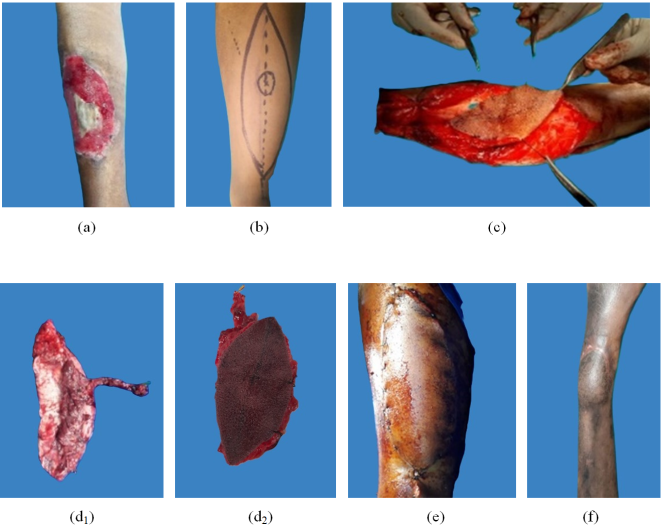
Figure 1. (a): Post-traumatic defect in the lower leg; 1 (b): Design of ALT flap; 1(c): Elevation of the flap; 1(d1 &d2): After harvesting of flap; 1(e): After inset of flap;; 1(f): After 1 year picture.
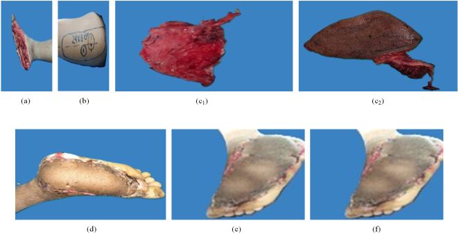
Figure 2. (a): Post-traumatic defect in the sole; 2 (b): Designing of ALT flap 2(c1 & c2): After harvest of flap; 2(d): At 8th POD View; 2(e): At 14th POD; 2(f): After 6months picture.
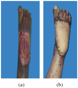
Figure 3. (a): Preoperative picture of the defect in the dorsum of the foot; (b): Post-operative picture.
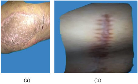
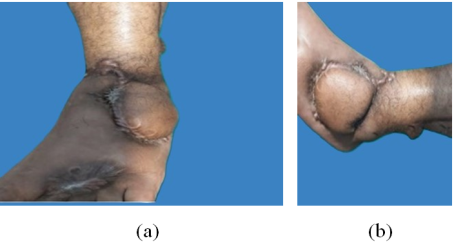
Figure 5. After 1-year follow-up picture.
Postoperative Care: Flap was monitored 4 hourly for the first 24 hours. Then 6 hourly in the next 24 hours and 12 hourly for another 48 hours for venous congestion or diminished arterial supply by checking color, temperature, capillary refill, turgor, and by hand-held doppler. We used Splint for two weeks. Then we did a 6-week night splint with physiotherapy at the time adequate. As of 1st POD antithrombotic prophylaxis with low molecular weight heparin subcutaneous enoxaparin and 100mg/day salicylic acid. Heparin should be continued for 3weeks and salicylic acid for six weeks.
Statistical Analysis: All data were recorded systematically in a preformed data collection form. Quantitative data was expressed as mean and standard deviation and qualitative data was expressed as frequency distribution and percentage. Statistical analysis was performed using SPSS 23 (Statistical Package for Social Sciences) for Windows version 10. The Ethical Review Committee of Rangpur Medical College Hospital approved the study.
3. Results
Table 1. Distribution of the respondents by demographic variables.
Demographic variables | N | P (%) |
Age | |
< 20 years | 2 | 12.5 |
20-29 years | 5 | 31.25 |
30-39 years | 3 | 18.75 |
>39 years | 6 | 37.5 |
Mean ±SD (years) | 34.25 ±15.27 |
Gender | |
Male | 12 | 75 |
Female | 4 | 25 |
Smoking status | | |
Yes | 6 | 37.5 |
No | 10 | 62.5 |
Defect origin | | |
Trauma | 13 | 81.25 |
Marjolin ulcer | 1 | 6.25 |
Electric burn | 2 | 12.5 |
Location of defect | | |
Sole & Heel (left/right) | 5 | 31.25 |
Dorsum of foot (left /right) | 4 | 25 |
The middle part of leg | 1 | 6.25 |
Lower 1/3rd of rt leg | 6 | 37.5 |
Mean Size (length, cm) | 6.625 ±1.902 |
Mean Size (wide, cm) | 3.868 ± 0.994 |
Mean Time delay (days) | 17.125 ± 5.760 |
Co-morbidities | | |
DM | 3 | 18.75 |
Hypothyroidism | 1 | 6.25 |
Among 16 patients, the majority (31.25%) of patients were aged 20-29 years and the mean age was 34.25 ±15.27 years. Twelve (75%) patients were males and 4 (25%) were females. The male and female ratio was 3:1 in our study. Among them 13 were traumatic cases, 2 were electrical burn wounds, and one was an oncologic case. Most (37.5%) of patients had defects in 1/3rd lower of right leg, followed by 31.25% had defects in heel areas. The mean delayed time was 17.125 ± 5.760 days. Among our respondents, 3 patients had DM and only 1 patient had hypothyroidism.
Table 2. Distribution of our study patients by intraoperative variables.
Intraoperative variables | N | P (%) |
Type of flap | |
Fascio cutaneous | 12 | 75.0 |
Fat cutaneous | 4 | 25.0 |
Number of perforating arteries | | |
1 | 13 | 81.3 |
2 | 3 | 19.7 |
Type of perforating arteries | | |
Septal | 4 | 25.0 |
Muscular | 12 | 75.0 |
Recipient vascular bundle | | |
Posterior tibial vessels | 4 | 25.0 |
Anterior tibial vessels | 12 | 75.0 |
Stitches | | |
8/o prolene | 16 | 100.0 |
Arterial anastomosis | | |
End to end | 14 | 87.5 |
End to side | 2 | 12.5 |
Venous anastomosis | | |
End to end | 16 | 100.0 |
Donor site closure | | |
Primary | 12 | 75.0 |
STSG | 4 | 25.0 |
The majority (75%) of patients had fascia cutaneous and the maximum (81.3%) surgeries were done on the first perforating artery and the most common perforating artery was muscular (75%). Anterior tibial vessels were done in 75% of patients. Among all patients, end-to-end arterial anastomosis was done in 87.5% while end-to-end venous anastomosis was done in all (100%) patients. Survival rates of the free flap were 87.5% (14) with two failure cases. There were no complications at the donor site. Primary closure was used in 12 cases whereas in four cases we used STSG.
Table 3. Outcome measurement scale.
Result | Flap survival rate | Donor site |
Excellent | Flap completely survived No venous congestion or arterial insufficiency, no infection, and excellent flap adhesion. | No infection, no wound dehiscence, closed primarily. |
Satisfactory | Flap survives with few complications. | Mild infection, Wound dehiscence, secondary closure |
Poor | Flap failure or loss, severe complications | Primary, Secondary closure, or STSG |
Table 3 shows the outcome measurement scale of our study respondents. The outcome of our study patients was divided into 3 scales – excellent (flap survived with no infection & no complications), satisfactory (flap survived with no complications), and poor (flap failure with severe complications).
Table 4. Distribution of patient by outcome.
Outcome | N | P (%) |
Excellent | 12 | 75 |
Satisfactory | 2 | 12.5 |
Poor | 2 | 12.5 |
In this study, 12(75%) cases showed excellent outcomes where there were no signs, or symptoms of venous congestion or arterial insufficiency, no need for exploration, no infection or dehiscence, and the donor site was primarily closed with good flap adhesion. There were 2 (12.5%) cases with satisfactory outcomes and venous congestion was present in those two patients. Only 2 cases had poor outcomes with severe complications and complete flap loss.
4. Discussion
The evaluation of the optimum lower extremity reconstruction approach may be challenging due to the different local tissue needs related to thickness, weight-bearing status, and shear stresses. The ALT flap is an excellent fit for this application due to its versatility in terms of both use and harvesting techniques. In this study, we classified reconstructions based on the exact anatomic location of the defect to better describe the applications of the ALT flap in lower extremity reconstruction. For anomalies in the hip and buttock region, local tissue reorganization or local flaps are usually adequate because of the region's abundance of soft tissues. When defects are more extensive or prior radiotherapy precludes the use of local tissue, an ipsilateral pedicled ALT flap with part of the vastus lateralis muscle can provide reliable soft tissue coverage, as shown in this series
[3] | Nosrati N, Chao Ah, Chang Dw, Yu P. Lower Extremity Reconstruction With The Anterolateral Thigh Flap. Journal Of Reconstructive Microsurgery. 2012 May; 28(04): 227-34. |
[3]
.
There are many reconstructive options available for upper & middle part of lower limb reconstruction. However, lower 1/3rd and foot reconstruction are unique & difficult as very few local options are available. Soft tissue reconstruction requires good vascularity, durability & adequate tissue volume. A well-padded flap is often necessary for heel and foot coverage.
Road traffic accidents are one of the major causes of mortality and morbidity and mostly affect young people, lower legs & feet suffer most. Cross-leg flaps were commonly used in the past. VAC therapy, skin graft, and local flap were also popular. In the beginning of the 20th century tube flaps were one of the reconstructive tools for leg & foot defects.
In advance of the development of medical science & lack of availability of local tissue & regional tissue, free tissue transfer has become popular in this context.
In this study, we operated 16 cases with an average age of 32 years (range 12-65). Active males were predominant to females with a ratio of 14:1.
Free ALT flaps have emerged as versatile options for lower leg and foot reconstructions due to their abundant blood supply, the edge of harvesting, and reliable outcome. The ALT flap provides ample tissue coverage, making it ideal for large tissue defects as proven in our clinical care
[18] | Nerlich AG, Zink A, Szeimies U, Hagedorn HG. Ancient Egyptian prosthesis of the big toe. The Lancet. 2000 Dec 23; 356(9248): 2176-9. |
[19] | Park Je, Rodriguez Ed, Bluebond-Langer R, Et Al. The Anterolateral Thigh Flap Is Highly Effective For Reconstruction Of Complex Lower Extremity Trauma. J Trauma. 2007; 62: 162-165. |
[20] | Jeng Sf, Wei Fc. Classification And Reconstructive Options In Foot Plantar Skin Avulsion Injuries. Plast Reconstr Surg 1997; 99: 1695-1705. |
[21] | Kohli Js, Pande S, Bajaja Sp. Large Transversefasciocutaneous Leg Flap: Whole Leg Flap In Prosess Citation. Br J Plast Surg 2000; 53: 495-8. |
[18-21]
. The ability to transfer vascularized tissue as a fascio cutaneous flap-sparing muscle also reduces the risk of donor site morbidity. Several studies have reported favorable outcomes of ALT-free flap in lower limb reconstruction
[22] | Rahim S. Nazerali, Lee L. Q. Pu. Free Tissue Transfer To Thelower Extrimity A Paradigm Shift In Flap Selection For Soft Tissue Reconstruction Ann Plast Surg 2013; 70: 419-422. |
[23] | Serafin D, Georgiade Ng, Smith Dh. Comparison Of Freeflaps With Pedicled Flaps For Coverage Of Defects Of The Leg Or Foot. Plast Reconstr Surg 2007; 59: 492-9. |
[24] | Gedebou Tm, Wei Fc, Lin Ch. Clinical Experience Of 1284 Free Anterolateral Thigh Flaps. Handchir Mikrochir Plast Chir 2002; 34(4): 239-44. |
[25] | Mureau Ma, Posch Na, Meeuwis Ca, Hofer So. Anterolateral Thigh Flap Reconstruction Of Large External Facial Skin Defects: A Follow-Up Study On Functional And Aesthetic Recipient- And Donor-Site Outcome. Plast Reconstr Surg 2005; 115(4): 1077-86. |
[22-25]
. In our study, we found 75% of cases were excellent, 12.5% were satisfactory, and 12.5% were poor. However, even when soft tissue coverage is achieved, functional recovery depends mainly on the recovery of the range of motion of the Ankle joint through tendon reconstruction. All of the patients who had their foot and ankle abnormalities fixed with ALT flaps returned to their preoperative functional status, which is consistent with previous research that demonstrated satisfactory functional status after limb salvage procedures for soft tissue sarcomas of the foot and ankle
[26] | Colterjohn Nr, Davis Am, O'sullivan B, Catton Cn, Wunder Js, Bell Rs. Functional Outcome In Limb-Salvage Surgery For Soft Tissue Tumours Of The Foot And Ankle. Sarcoma 1997; 1(2): 67–74. |
[26]
. Compared to more proximal portions of the leg, the foot and ankle require a thinner and more flexible replacement. The ALT flap works effectively for this in persons who are not fat
[9] | Demirtas Y, Neimetzade T, Kelahmetoglu O, Guneren E. Comparison Of Free Muscle And Perforator Skin Flaps For Soft Tissue Reconstruction Of The Foot And Ankle. Foot Ankle Int 2010; 31(1): 53–58.. |
[10] | Hong Jp, Kim Ek. Sole Reconstruction Using Anterolateral Thigh Perforator Free Flaps. Plast Reconstr Surg 2007; 119(1): 186–193. |
[9, 10]
. However, the ALT flap is frequently much thicker in Western nations, which restricts its application
[27] | Yu P. Characteristics Of The Anterolateral Thigh Flap In A Western Population And Its Application In Head And Neck Reconstruction. Head Neck 2004; 26(9): 759–769. |
[27]
. Flap thinning is feasible, but it won't produce a flap that is as thin and flexible as, say, a radial forearm flap.
5. Limitations of the Study
Our study was a single-center study. We took a small sample size due to our short study period. After evaluating those patients, we did not follow up with them for the long term and did not know other possible interference that may happen in the long term with these patients.
6. Conclusion and Recommendations
In our study, we found that the use of a free ALT flap was effective in meeting the needs of our young patients. The successful outcome of this study underlines the importance of further research & studies to explore the potential of combining different reconstructive methods to improve limb salvage rates.
So further study with a prospective and longitudinal study design including a larger sample size needs to be done to assess the functional, and aesthetic outcome of these procedures.
Abbreviations
ALT | Anterior Lateral Thigh Flap |
VL | Vastus Lateralis |
STSG | Split-Thickness Skin Graft |
COPD | Chronic Obstructive Pulmonary Disease |
Author Contributions
M. A. Hamid: Conceptualization, Data curation, Formal Analysis, Funding acquisition, Investigation, Methodology, Project administration, Resources, Software, Supervision, Validation, Visualization, Writing – original draft, Writing – review & editing
Sayeda Niger Sultana: Conceptualization, Funding acquisition, Methodology, Software, Visualization
Shah Md. Al Mukit: Funding acquisition, Investigation, Resources, Validation, Visualization, Writing – review & editing
Goutam Biswas: Project administration Resources Supervision Writing – original draft
Funding
No funding sources.
Ethical Approval
The study was approved by the Institutional Ethics Committee.
Conflict of Interest
The authors declare no conflicts of interest.
References
[1] |
Song Yg, Chen Gz, Song Yl. The Free Thigh Flap: A New Free Flap Based On The Septocutaneous Artery. Br J Plast Surg 1984; 37: 149–159.
|
[2] |
Mardini S, Lin Ch, Wei Fc. In: Blondeel Pn, Morris Sf, Hallock Gg, Et Al, Eds. Perforator Flaps: Anatomy, Technique & Clinical Applications. St. Louis, Mo: Quality Medical Publishing; 2006: 617–633.
|
[3] |
Nosrati N, Chao Ah, Chang Dw, Yu P. Lower Extremity Reconstruction With The Anterolateral Thigh Flap. Journal Of Reconstructive Microsurgery. 2012 May; 28(04): 227-34.
|
[4] |
Lee N, Roh S, Yang K, Kim J. Reconstruction Of Hand And Forearm After Sarcoma Resection Using Anterolateral Thigh Free Flap. J Plast Reconstr Aesthet Surg 2009; 62(12): E584–E586.
|
[5] |
Gravvanis A, Tsoutsos D, Karakitsos D, Iconomou T, Papadopoulos O. Blood Perfusion Of The Free Anterolateral Thigh Perforator Flap: Its Beneficial Effect In The Reconstruction Of Infected Wounds In The Lower Extremity. World J Surg 2007; 31(1): 11–18.
|
[6] |
Kuo Yr, Kuo Mh, Chou Wc, Liu Yt, Lutz Bs, Jeng Sf. One-Stage Reconstruction Of Soft Tissue And Achilles Tendon Defects Using A Composite Free Anterolateral Thigh Flap With Vascularized Fascia Lata: Clinical Experience And Functional Assessment. Ann Plast Surg 2003; 50(2): 149–155.
|
[7] |
Heller L, Kronowitz Sj. Lower Extremity Reconstruction. J Surg Oncol 2006; 94(6): 479–489.
|
[8] |
Park Je, Rodriguez Ed, Bluebond-Langer R, Et Al. The Anterolateral Thigh Flap Is Highly Effective For Reconstruction Of Complex Lower Extremity Trauma. J Trauma 2007; 62(1): 162–165.
|
[9] |
Demirtas Y, Neimetzade T, Kelahmetoglu O, Guneren E. Comparison Of Free Muscle And Perforator Skin Flaps For Soft Tissue Reconstruction Of The Foot And Ankle. Foot Ankle Int 2010; 31(1): 53–58..
|
[10] |
Hong Jp, Kim Ek. Sole Reconstruction Using Anterolateral Thigh Perforator Free Flaps. Plast Reconstr Surg 2007; 119(1): 186–193.
|
[11] |
Ng Rw, Chan Jy, Mok V, Li Gk. Clinical Use Of A Pedicled Anterolateral Thigh Flap. J Plast Reconstr Aesthet Surg 2008; 61(2): 158– 164.
|
[12] |
Friji Mt, Suri Mp, Shankhdhar Vk, Ahmad Qg, Yadav Ps. Pedicled Anterolateral Thigh Flap: A Versatile Flap For Difficult Regional Soft Tissue Reconstruction. Ann Plast Surg 2010; 64(4): 458–461.
|
[13] |
Gravvanis Ai, Tsoutsos Da, Karakitsos D, Et Al. Application Of The Pedicled Anterolateral Thigh Flap To Defects From The Pelvis To The Knee. Microsurgery 2006; 26(6): 432–438.
|
[14] |
Wei Fc, Jain V, Celik N, Chen Hc, Chuang Dc, Lin Ch. Have We Found An Ideal Soft-Tissue Flap? An Experience With 672 Anterolateral Thigh Flaps. Plastic And Reconstructive Surgery. 2002 Jun 1; 109(7): 2219-26.
|
[15] |
Biswas D, Kalam Ma, Ahmed T, Khan Mrk. Results Of Microvascular Free Flap For Reconstruction Of Soft Tissue Defects: Our Early Experience. Bdjps 2012; 3(2): 33-37.
|
[16] |
Michael R. Christy, Avron Lipschitz, Eduardo Rodriguez, Karan Chopra, Nance Yuan, Early Postoperative Outcomes Associated With The Anterolateral Thigh Flap In Gustiloiiib Fractures Of The Lower Extremity. Ann Plast Surg2014; 72: 80-83.
|
[17] |
Hossein Nasajpour, Matthew H. Steele, Anterolateral Thighfree Flap For “Head-To-Toe” Reconstruction. Ann Plast Surg 2011; 66: 530–533.
|
[18] |
Nerlich AG, Zink A, Szeimies U, Hagedorn HG. Ancient Egyptian prosthesis of the big toe. The Lancet. 2000 Dec 23; 356(9248): 2176-9.
|
[19] |
Park Je, Rodriguez Ed, Bluebond-Langer R, Et Al. The Anterolateral Thigh Flap Is Highly Effective For Reconstruction Of Complex Lower Extremity Trauma. J Trauma. 2007; 62: 162-165.
|
[20] |
Jeng Sf, Wei Fc. Classification And Reconstructive Options In Foot Plantar Skin Avulsion Injuries. Plast Reconstr Surg 1997; 99: 1695-1705.
|
[21] |
Kohli Js, Pande S, Bajaja Sp. Large Transversefasciocutaneous Leg Flap: Whole Leg Flap In Prosess Citation. Br J Plast Surg 2000; 53: 495-8.
|
[22] |
Rahim S. Nazerali, Lee L. Q. Pu. Free Tissue Transfer To Thelower Extrimity A Paradigm Shift In Flap Selection For Soft Tissue Reconstruction Ann Plast Surg 2013; 70: 419-422.
|
[23] |
Serafin D, Georgiade Ng, Smith Dh. Comparison Of Freeflaps With Pedicled Flaps For Coverage Of Defects Of The Leg Or Foot. Plast Reconstr Surg 2007; 59: 492-9.
|
[24] |
Gedebou Tm, Wei Fc, Lin Ch. Clinical Experience Of 1284 Free Anterolateral Thigh Flaps. Handchir Mikrochir Plast Chir 2002; 34(4): 239-44.
|
[25] |
Mureau Ma, Posch Na, Meeuwis Ca, Hofer So. Anterolateral Thigh Flap Reconstruction Of Large External Facial Skin Defects: A Follow-Up Study On Functional And Aesthetic Recipient- And Donor-Site Outcome. Plast Reconstr Surg 2005; 115(4): 1077-86.
|
[26] |
Colterjohn Nr, Davis Am, O'sullivan B, Catton Cn, Wunder Js, Bell Rs. Functional Outcome In Limb-Salvage Surgery For Soft Tissue Tumours Of The Foot And Ankle. Sarcoma 1997; 1(2): 67–74.
|
[27] |
Yu P. Characteristics Of The Anterolateral Thigh Flap In A Western Population And Its Application In Head And Neck Reconstruction. Head Neck 2004; 26(9): 759–769.
|
Cite This Article
-
APA Style
Hamid, M. A., Sultana, S. N., Mukit, S. M. A., Biswas, G. (2024). The Anterolateral Thigh Flap for Lower Leg & Foot Reconstruction in a Tertiary Hospital in Northern Bangladesh: Our Observation and Outcome. American Journal of Health Research, 12(6), 230-236. https://doi.org/10.11648/j.ajhr.20241206.17
Copy
|
Download
ACS Style
Hamid, M. A.; Sultana, S. N.; Mukit, S. M. A.; Biswas, G. The Anterolateral Thigh Flap for Lower Leg & Foot Reconstruction in a Tertiary Hospital in Northern Bangladesh: Our Observation and Outcome. Am. J. Health Res. 2024, 12(6), 230-236. doi: 10.11648/j.ajhr.20241206.17
Copy
|
Download
AMA Style
Hamid MA, Sultana SN, Mukit SMA, Biswas G. The Anterolateral Thigh Flap for Lower Leg & Foot Reconstruction in a Tertiary Hospital in Northern Bangladesh: Our Observation and Outcome. Am J Health Res. 2024;12(6):230-236. doi: 10.11648/j.ajhr.20241206.17
Copy
|
Download
-
@article{10.11648/j.ajhr.20241206.17,
author = {M. A. Hamid and Sayeda Niger Sultana and Shah Md. Al Mukit and Goutam Biswas},
title = {The Anterolateral Thigh Flap for Lower Leg & Foot Reconstruction in a Tertiary Hospital in Northern Bangladesh: Our Observation and Outcome
},
journal = {American Journal of Health Research},
volume = {12},
number = {6},
pages = {230-236},
doi = {10.11648/j.ajhr.20241206.17},
url = {https://doi.org/10.11648/j.ajhr.20241206.17},
eprint = {https://article.sciencepublishinggroup.com/pdf/10.11648.j.ajhr.20241206.17},
abstract = {Introduction: The Anterior Lateral thigh flap (ALT) free flap can be considered one of the reconstructive tools in lower leg and foot reconstruction. It has a long and reliable pedicle, and dissection is comparatively easy, which makes it extremely suitable as a free flap. This study aimed to evaluate the versatility of the flap in covering lower leg and foot defects. Methods: This was a prospective observational study conducted in the Department of Plastic Surgery, Rangpur Medical College Hospital, Rangpur, Bangladesh, from January 2023 to December 2023. In our study, we included 16 patients with defects in the lower leg and foot who received an ALT-free flap as a reconstructive treatment. We analyzed the preoperative, intraoperative, and postoperative variables of our respondents. Results: In this study, 16 patients (12 males and 4 females) with an average of 34.25 years old were included. Among them 13 were traumatic cases, 2 electrical burn wounds, and one oncologic case. Survival rates of the free flap were 87.5% (14) with two failure cases. There were no complications at the donor site. In this study, 75% (12) cases showed excellent outcomes where there were no signs, or symptoms of venous congestion or arterial insufficiency, no infection or dehiscence, and the donor site was primarily closed with good flap adhesion. Conclusion: This study shows that the ALT-free flap is a versatile flap that allows it to cover the defect successfully and ALT adaptation was satisfactory in terms of varied location and size.
},
year = {2024}
}
Copy
|
Download
-
TY - JOUR
T1 - The Anterolateral Thigh Flap for Lower Leg & Foot Reconstruction in a Tertiary Hospital in Northern Bangladesh: Our Observation and Outcome
AU - M. A. Hamid
AU - Sayeda Niger Sultana
AU - Shah Md. Al Mukit
AU - Goutam Biswas
Y1 - 2024/11/29
PY - 2024
N1 - https://doi.org/10.11648/j.ajhr.20241206.17
DO - 10.11648/j.ajhr.20241206.17
T2 - American Journal of Health Research
JF - American Journal of Health Research
JO - American Journal of Health Research
SP - 230
EP - 236
PB - Science Publishing Group
SN - 2330-8796
UR - https://doi.org/10.11648/j.ajhr.20241206.17
AB - Introduction: The Anterior Lateral thigh flap (ALT) free flap can be considered one of the reconstructive tools in lower leg and foot reconstruction. It has a long and reliable pedicle, and dissection is comparatively easy, which makes it extremely suitable as a free flap. This study aimed to evaluate the versatility of the flap in covering lower leg and foot defects. Methods: This was a prospective observational study conducted in the Department of Plastic Surgery, Rangpur Medical College Hospital, Rangpur, Bangladesh, from January 2023 to December 2023. In our study, we included 16 patients with defects in the lower leg and foot who received an ALT-free flap as a reconstructive treatment. We analyzed the preoperative, intraoperative, and postoperative variables of our respondents. Results: In this study, 16 patients (12 males and 4 females) with an average of 34.25 years old were included. Among them 13 were traumatic cases, 2 electrical burn wounds, and one oncologic case. Survival rates of the free flap were 87.5% (14) with two failure cases. There were no complications at the donor site. In this study, 75% (12) cases showed excellent outcomes where there were no signs, or symptoms of venous congestion or arterial insufficiency, no infection or dehiscence, and the donor site was primarily closed with good flap adhesion. Conclusion: This study shows that the ALT-free flap is a versatile flap that allows it to cover the defect successfully and ALT adaptation was satisfactory in terms of varied location and size.
VL - 12
IS - 6
ER -
Copy
|
Download