Persistent Epstein Baar Virus (EBV) infection may be a perfect target for the treatment of EBV-associated lymphomas and improved patient outcomes. The aim of this work was to evaluate the frequency of LMP1 (Latence membrane Protein 1) expression in pediatric lymphomas in Dakar. Material and Methods: This was a retrospective, descriptive study from January 1, 2015 to December 31, 2020. It was based on blocks and anatomopathological reports of pediatric Lymphomas in 04 ACP laboratories in Dakar. The immunohistochemical study was carried out at the IBN ROCHD Hospital in Casablanca, Morocco, using a manual method. LMP1 immunostaining was considered positive if 10% of tumour cells showed cytoplasmic staining. Results: Positive staining for LMP1 was noted in 13 of the 42 cases of pediatric lymphomas tested, i.e. 31% of cases. These included 09 cases of Hodgkin's lymphoma, i.e. 69% (9/13), and 04 cases of non-Hodgkin's lymphoma, i.e. 31%. LMP1-positive classical Hodgkin's lymphomas accounted for 40.09% (9/22) of Hodgkin's lymphomas, and were of the mixed cellularity (6/22) and scleronodular (3/22) subtypes. The frequency of LMP1-positive samples in non-Hodgkin's lymphomas was 20% (4/20). These included 3 cases of Burkitt's lymphoma and 1 case of diffuse large B-cell lymphoma. Conclusion: The frequency of EBV infection in pediatric lymphomas in Senegal is lower than in endemic areas of Africa.
This is an Open Access article, distributed under the terms of the Creative Commons Attribution 4.0 International License (http://creativecommons.org/licenses/by/4.0/), which permits unrestricted use, distribution and reproduction in any medium or format, provided the original work is properly cited.
Epstein-Barr virus (EBV) is a double-stranded DNA virus belonging to the Herpes viridae family. It was isolated from Burkitt's lymphoma (BL) cells almost 50 years ago (1964)
[1]
Epstein MA, Achong BG, Barr YM. Virus particles in cultured lymphoblasts from Burkitt’s lymphoma. Lancet. 1964; 1(7335): 702–3.
. EBV is the most widespread virus in humans, present in all geographic regions and infecting over 90% of the world's population. In developing countries, infections occur very early in life, causing no specific features other than the general symptoms of acute viremia. In developed countries, however, infection is generally delayed until adolescence or early adulthood, when it causes a benign, spontaneous lymphoproliferative disorder known as infectious mononucleosis
The virus in target cells expresses six nuclear antigens (EBNA1, 2, 3A, 3B, 3C and LP), two small monocoding RNAs (EBER1 and 2) and three latent membrane proteins (LMP1, 2A and 2B)
[3]
Rickinson A. Epstein-Barr virus. Virus Res. 2002; 82(1-2): 109–13.
Epstein-Barr virus may be responsible for a number of B-cell lymphoproliferative disorders, such as classical Hodgkin's lymphoma (CHL) and Burkitt's lymphoma. In addition, this virus may have an association with a group of T-cell lymphomas of peripheral origin, such as nasal natural killer/extranodal T-cell lymphomas, angioimmunoblastic T-cell lymphomas, gastric adenocarcinoma and smooth muscle tumors
[4]
Hashemian M, Shirvani F. Epstein-Barr Virus-associated Pediatric Neoplasms. Arch Pediatr Infect Dis. 2020; 8(1): e94371.
. The frequency of this link varies both between and within countries. It also varies according to lymphoma histological subtype, gender and age at diagnosis.
In Senegal, the association between EBV and undifferentiated nasopharyngeal carcinomas has been studied
[6]
Gaye AM, Mouamba FG, Diemé MJ, Niang D, Thiam I, Dial CMM. Undifferentiated carcinomas of the nasopharynx in Dakar, a supposedly non-endemic area: about 13 cases. Bull. Soc. Pathol. Exot. 2018; 111: 84-89.
. However, few studies have focused on the relationship between lymphomas and Epstein-Barr virus.
The aim of this work was to evaluate the frequency of LMP1 expression in pediatric lymphomas in Dakar and its correlation with clinico-histological parameters.
2. Material and Methods
This is a retrospective, descriptive study of a series of cases over six (06) years, from January 1, 2015 to December 31, 2020.
It was conducted in the pathological anatomy laboratories of 04 national hospitals in Dakar (Hôpital Aristide Le Dantec, Hôpital Général Idrissa POUYE, Hôpital de FANN and Hôpital Principal de Dakar), where 42 kerosene blocks of pediatric lymphoma cases were recorded.
Patients fulfilling the following conditions were included in this study:
1) Age < 18 years at diagnosis.
2) All cases of Hodgkin lymphoma (HL) and non-Hodgkin lymphoma (NHL).
3) Diagnoses based on histology and immunohistochemistry (cytologies often used constitute a major bias).
4) All sites included.
All cases were reviewed by two senior pathologists and confirmed by immunohistochemistry.
LMP1 immunohistochemistry was performed at the pathological anatomy laboratory of Ibn ROCHD Hospital (Casablanca).
The manual method was used for LMP1 immunohistochemistry (clone: CS.1-4, batch: 41247654).
LMP1 immunostaining was considered positive if 10% of tumour cells showed cytoplasmic staining. LMP1 expression was correlated with clinicopathological parameters. The correlation was statistically significant if p ≤ 0.05.
Data were analyzed using Excel 2010 and SPSS20.0.
3. Results
Positive staining was noted in 13 of the 42 cases of pediatric lymphomas tested, i.e. 31%.
These included 09 cases of Hodgkin's lymphoma, i.e. 69% (9/13), and 04 cases of non-Hodgkin's lymphoma, i.e. 31% (4/13).
Classical LMP1-positive Hodgkin's lymphomas accounted for 40.09% (9/22) of CHL, with mixed cellularity (6/22) and sclerodular subtypes (3/22). The mean age of CHL patients with LMP1 was 11.56 years. All were lymph node positive and 88.9% were male. Localized stages accounted for 66.7% of LMP1-positive CHL versus 33.3% for advanced stages.
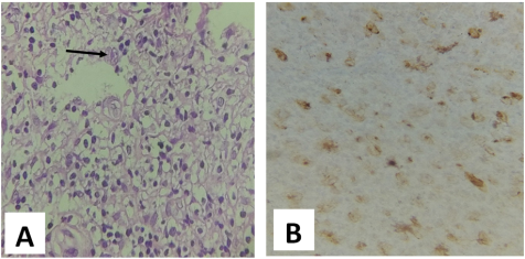
The LMP1 positivity rate was 54.54% (6/11) in mixed cellularity CHL (figure 1) and 30% (3/10) in sclerodular type CHL.
The correlation between clinico-pathological parameters of Hodgkin's lymphoma and LMP1 expression was statistically insignificant (Table 1).
The frequency of LMP1-positive samples in NHL was 20% (4/20). LMP1-positive NHLs were all of B phenotype. These included 3 cases of Burkitt's lymphoma (BL) and 1 case of diffuse large B-cell lymphoma (DLBCL).
The mean age of patients with LMP1-positive NHL was 10.25 years. All were male and lymph node positive. LMP1 expression was found in favorable stages (I and II) in one case and in unfavorable stages (III and IV) in 3 cases.
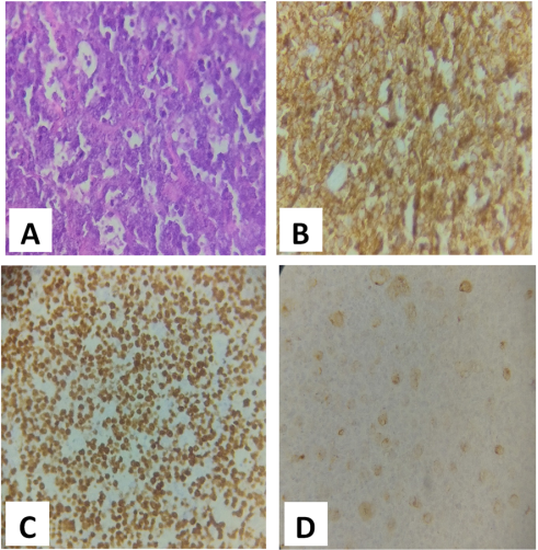
LMP1-positive Burkitt's lymphomas accounted for 50% (3/6) of Burkitt's lymphomas (figure 2).
LMP1-positive diffuse large B-cell lymphoma accounted for 10% (1/10) of DLBCL (figure 3). No statistically significant relationship was observed between LMP1 expression and clinico-pathological parameters of NHL (table 2).
Table 1. Correlation between clinico-pathological parameters of Hodgkin's lymphoma and LMP1 expression (n=22).
LMP1 positive (n=9)
LMP1 négative (n=13)
Total (n=22)
P value
Age (ans)
0,823
Mean Age
11,56 ±3,6
11,92 ±3,8
11,77 ± 3,6
Median Age
12 (5-15)
13 (5-16)
12,50 (5-16)
Sexe
0,157
Male
8 (88,9%)
8 (61,5%)
16
Female
1 (11,1%)
5 (38,5%)
6
Siège
0,217
Ganglionar
9 (100%)
11 (86,4%)
20
Extraganglionar
0 (00%)
2 (15,4%)
2
Clinical stages
0,806
I et II
6 (66,7%)
8 (61,5%)
14
III et IV
3 (33,3)
5 (38,5)
8
Histological subtypes
0,518
Scleronodular
3 (33,3)
6 (46,2%)
9
Mixed cellularity
6 (66,7%)
6 (46,2%)
12
Rich in lymphocytes
0 (00%)
1 (7,7%)
1
Table 2. Corrélation entre paramètres clinico-pathologiques des Lymphomes non Hodgkiniens et le statut de LMP1 (n=20).
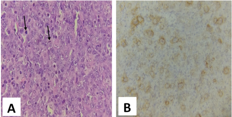
Figure 3. Microscopic appearance of diffuse large B-cell lymphoma expressing LMP1.
A: Tumour proliferation of large atypical cells in sheets with mitoses (arrow) HE x 400.
B: Cytoplasmic staining of tumour cells with anti-LMP1 antibody (X 400 immunohistochemistry).
4. Discussion
Major progress has been made in understanding the pathological features of lymphoid neoplasms in children and adolescents over the past 5 years, leading to the WHO's broadening of the spectrum of EBV-related lymphomas.
The frequency of association between lymphoma and EBV varies both between and within countries. It also varies according to lymphoma histological subtype, gender and age at diagnosis.
The rate of LMP1-positive pediatric lymphomas after immunohistochemistry was 31% (13/42). This result is similar to that reported by Osman et al at the South Egypt Cancer Institute with a frequency of 32.02% (25/78)
[7]
Osman AM, Ali AM, Shaban SH, Helmy EM, Badrawy H, Shibl A. Detection of Epstein-Barr virus Latent Membrane Protein-1 in Pediatric Lymphoma: A Report from South Egypt Cancer Institute. SECI Oncology 2022; (2): 101-107.
[7]
, and significantly lower than the rate observed in Iran with 62.5%
[8]
Zareifar S, Kazemi B, Arzanian MT, Bandehpour M. Detection of Epstein–Barr virus in Pediatric Lymphoma: A Single Center Study. J Leuk 2016; 4: 213.
The frequency of this association will make it possible to encourage research projects for a vaccine targeting EBV, and to include it in vaccination programs for children from birth in Senegal, as has been the case for hepatitis B and human papillomavirus (HPV).
LMP-1 positivity in HL tumor samples in this study (40.09%), is close to the 38.5% observed by Osman et al
[7]
Osman AM, Ali AM, Shaban SH, Helmy EM, Badrawy H, Shibl A. Detection of Epstein-Barr virus Latent Membrane Protein-1 in Pediatric Lymphoma: A Report from South Egypt Cancer Institute. SECI Oncology 2022; (2): 101-107.
[7]
. This association is much lower than that noted in South Africa and Jordan, where rates were 68%
[9]
Engel M, Essop MF, Close P, Hartley P, Pallesen G, Sinclair-Smith C. Improved prognosis of Epstein-Barr virus associated childhood Hodgkin’s lymphoma: study of 47 South African cases. J Clin Pathol 2000; 53: 182–186.
. The involvement of EBV in the occurrence of CHL varies according to geographical region and socio-economic level. In the meta-analysis by Lee et al, EBV positivity of over 70% was found in pediatric CHL in Africa, Asia and Central and South America, whereas in Europe and North America the rate was lower, around 40%
[11]
Lee JH, Kim Y, Choi JW, Kim YS. Prevalence and prognostic significance of Epstein-Barr virus infection in classical Hodgkin's lymphoma: a meta-analysis. Arch Med Res. 2014; 45(5): 417-31.
The male predominance and exclusive lymph node localization noted in this study were also reported by other authors
[7]
Osman AM, Ali AM, Shaban SH, Helmy EM, Badrawy H, Shibl A. Detection of Epstein-Barr virus Latent Membrane Protein-1 in Pediatric Lymphoma: A Report from South Egypt Cancer Institute. SECI Oncology 2022; (2): 101-107.
[12]
Qin C, Huang Y, Feng Y, Li M, Guo N, Rao H. Clinicopathological features and EBV infection status of lymphoma in children and adolescents in South China: a retrospective study of 662 cases. Diagnostic Pathology 2018; 13: 17.
. In our series, however, the mean age was 11.55 years.
The frequency of EBV-associated HL was closer to that observed in developed countries. This lower incidence of EBV-positive HL in our series would appear to be linked, on the one hand, to the absence of immunosuppression and, on the other, to the timing of EBV infection, which occurs a little later in life (mean age 11, 55 years), resulting in a lower risk of developing HL compared with individuals with a previous history.
EBV is detected in Hodgkin and Reed-Sternberg cells, mainly in mixed cellularity and lymphocyte-depleted subtypes, and less frequently in scleronodular and lymphocyte-rich subtypes
[11]
Lee JH, Kim Y, Choi JW, Kim YS. Prevalence and prognostic significance of Epstein-Barr virus infection in classical Hodgkin's lymphoma: a meta-analysis. Arch Med Res. 2014; 45(5): 417-31.
. In our cohort, the mixed cellularity subtype was more frequently associated with EBV, followed by the scleronodular subtype.
The correlation between LMP1 expression and clinical parameters in CHL was not significant. In the study by Osman et al
[7]
Osman AM, Ali AM, Shaban SH, Helmy EM, Badrawy H, Shibl A. Detection of Epstein-Barr virus Latent Membrane Protein-1 in Pediatric Lymphoma: A Report from South Egypt Cancer Institute. SECI Oncology 2022; (2): 101-107.
[7]
, only patient age was significantly associated with LMP1 status (p= 0.003).
Although numerous studies have reported a strong association between EBV-positive cases and the prevalence of the mixed cellularity subtype, reaching high frequencies of up to 94%
[14]
Araujo I, Bittencourt AL, Barbosa HS, et al. The high frequency of EBV infection in pediatric Hodgkin lymphoma is related to the classical type in Bahia, Brazil. Virchows Arch. 2006; 449(3): 315-9.
, the LMP1-positive cases in our study showed no significant difference in histological subtype distribution, which is similar to what was reported by Barros et al
[15]
Barros MHM, Hassan R, Niedobitek G. Tumorassociated macrophages in pediatric classical Hodgkin lymphoma: Association with EpsteinBarr virus, lymphocyte subsets, and prognostic impact. Clin Cancer Res. 2012; 18(14): 3762-71.
LMP1 expression in CHL was 66.7% for early stages (I and II) versus 33.3% for late stages (III and IV). This greater expression of LMP1 in early-stage HL has also been reported in other studies
[7]
Osman AM, Ali AM, Shaban SH, Helmy EM, Badrawy H, Shibl A. Detection of Epstein-Barr virus Latent Membrane Protein-1 in Pediatric Lymphoma: A Report from South Egypt Cancer Institute. SECI Oncology 2022; (2): 101-107.
[16]
Chabay PA, Mário H, Rocio DM, Elena R, Guadalupe C, Maria Z, et al. Pediatric Hodgkin Lymphoma in 2 South American Series: A Distinctive Epidemiologic Pattern and Lack of Association of Epstein-Barr Virus With Clinical Outcome. Journal of pediatric hematology/oncology 2008; 30: 285-91.
. However, no significant correlation was found between LMP1 expression and clinical stages.
The frequency of LMP1-positive samples in NHL was 20% in this study. This result was slightly higher than that of Peh et al, in Malaysia, where the association between childhood NHL and EBV was 18%
[17]
Peh SC, Madarajah VS, Tai YC, et al. Pattern of Epstein-Barr virus association in childhood nonHodgkin’s lymphoma: Experience of University of Malaya Medical Center. Pathol Int. 2004; 54(3): 151-7.
Other authors had observed frequencies significantly higher, with positivity rates of 28.8% and 65.85%
[7]
Osman AM, Ali AM, Shaban SH, Helmy EM, Badrawy H, Shibl A. Detection of Epstein-Barr virus Latent Membrane Protein-1 in Pediatric Lymphoma: A Report from South Egypt Cancer Institute. SECI Oncology 2022; (2): 101-107.
[8]
Zareifar S, Kazemi B, Arzanian MT, Bandehpour M. Detection of Epstein–Barr virus in Pediatric Lymphoma: A Single Center Study. J Leuk 2016; 4: 213.
The association of EBV with NHL varied according to age and sex, with a slightly greater distribution in boys over 11 years of age. However, there was no statistically significant association. In other studies, a significant association was noted in boys under 5 years of age
[8]
Zareifar S, Kazemi B, Arzanian MT, Bandehpour M. Detection of Epstein–Barr virus in Pediatric Lymphoma: A Single Center Study. J Leuk 2016; 4: 213.
The EBV-related NHL (LMP1-positive) were all B-phenotype lymphomas, corresponding to a positivity rate of 22.22% (4/18) of B-type NHL. Peh et al
[17]
Peh SC, Madarajah VS, Tai YC, et al. Pattern of Epstein-Barr virus association in childhood nonHodgkin’s lymphoma: Experience of University of Malaya Medical Center. Pathol Int. 2004; 54(3): 151-7.
. One of the most striking features of pediatric BL is the variation in the frequency of EBV positivity in different geographical regions.
The "high incidence" endemic BL found in equatorial Africa and New Guinea is around 95% EBV positive. Sporadic BL varies from 20% to 80% EBV seropositivity depending on geographic region, with the lowest viral association observed in Western countries
[19]
Hutcheson RL, Chakravorty A, Sugden B. Burkitt Lymphomas Evolve to Escape Dependencies on Epstein-Barr Virus. Front Cell Infect Microbiol. 2021; 10: 1–15.
HIV-associated BL was very different in pediatrics, where an association with EBV of interest of 100% was found, higher than the typical 30-40% reported in adults
[13]
Chabay P, Preciado MV. Epidemiology of Epstein-Barr virus-associated pediatric lymphomas from Argentina. Bol Med Hosp Infant Mex 2016; 73: 47-54.
Ismail A, Osman I, Husain NE. LMP1 Immunohistochemistry in Non-Hodgkin’s Lymphoma of Sudanese Cases. Open J Pathol. 2016; 06(02): 79-87. Open Journal of Pathology, 2016, 6, 79-87.
The pediatric form of LMP-1-positive DLBCL shows marked geographical differences that are profoundly influenced by socio-economic conditions.
Sporadic cases have been described in some Asian, African and Latin American countries, whereas they are extremely rare in Western countries and are generally associated with immunodeficiency
[21]
Uccini S, Al-Jadiry MF, Scarpino S, et al. Epstein Barr virus-positive diffuse large B-cell lymphoma in children: A disease reminiscent of Epstein-Barr virus-positive diffuse large B-cell lymphoma of the elderly. Hum Pathol. 2015; 46(5): 716-24.
In this study, 10% (1/10) of DLBCL specimens had an association with EBV, which is lower than observations reported by other authors
[7]
Osman AM, Ali AM, Shaban SH, Helmy EM, Badrawy H, Shibl A. Detection of Epstein-Barr virus Latent Membrane Protein-1 in Pediatric Lymphoma: A Report from South Egypt Cancer Institute. SECI Oncology 2022; (2): 101-107.
[12]
Qin C, Huang Y, Feng Y, Li M, Guo N, Rao H. Clinicopathological features and EBV infection status of lymphoma in children and adolescents in South China: a retrospective study of 662 cases. Diagnostic Pathology 2018; 13: 17.
The two cases of DLBCL occurring in immunocompromised children were not positive for LMP1.
LMP1 expression in NHL was more frequent in late stages, both in our series and in that of Osman et al
[7]
Osman AM, Ali AM, Shaban SH, Helmy EM, Badrawy H, Shibl A. Detection of Epstein-Barr virus Latent Membrane Protein-1 in Pediatric Lymphoma: A Report from South Egypt Cancer Institute. SECI Oncology 2022; (2): 101-107.
[7]
. On the other hand, in the cohort of Chabay et al
[16]
Chabay PA, Mário H, Rocio DM, Elena R, Guadalupe C, Maria Z, et al. Pediatric Hodgkin Lymphoma in 2 South American Series: A Distinctive Epidemiologic Pattern and Lack of Association of Epstein-Barr Virus With Clinical Outcome. Journal of pediatric hematology/oncology 2008; 30: 285-91.
No significant relationship was found between LMP1 expression and clinico-pathological parameters in the NHLs in this study. Osman et al
[7]
Osman AM, Ali AM, Shaban SH, Helmy EM, Badrawy H, Shibl A. Detection of Epstein-Barr virus Latent Membrane Protein-1 in Pediatric Lymphoma: A Report from South Egypt Cancer Institute. SECI Oncology 2022; (2): 101-107.
[7]
, had observed a significant correlation between LMP1 expression and age, sex and histological subtypes of NHL.
5. Conclusion
In Senegal, the association between EBV and pediatric lymphomas exists with a non-negligible frequency (31%). This link can be assessed using automated or manual immunohistochemistry of the anti-LMP1 antibody. The hypothesis that DLBCL occurring in HIV+ subjects are associated with EBV in 100% of cases has not been confirmed in this work.
Abbreviations
EBV: Epstein Baar Virus
LB: Burkitt Lymphoma
CHL: Classic Hodgkin Lymphoma
HL: Hodgkin Lymphoma
NHL: Non Hodgkin Lymphoma
DLBCL: Diffuse Large B- Cell Lymphoma
WHO: World Health Organization
HPV: Human Papilloma Virus
HIV: Human Immuodefiency Virus
Conflicts of Interest
The authors declare no conflicts of interest.
References
[1]
Epstein MA, Achong BG, Barr YM. Virus particles in cultured lymphoblasts from Burkitt’s lymphoma. Lancet. 1964; 1(7335): 702–3.
Osman AM, Ali AM, Shaban SH, Helmy EM, Badrawy H, Shibl A. Detection of Epstein-Barr virus Latent Membrane Protein-1 in Pediatric Lymphoma: A Report from South Egypt Cancer Institute. SECI Oncology 2022; (2): 101-107.
[8]
Zareifar S, Kazemi B, Arzanian MT, Bandehpour M. Detection of Epstein–Barr virus in Pediatric Lymphoma: A Single Center Study. J Leuk 2016; 4: 213.
Engel M, Essop MF, Close P, Hartley P, Pallesen G, Sinclair-Smith C. Improved prognosis of Epstein-Barr virus associated childhood Hodgkin’s lymphoma: study of 47 South African cases. J Clin Pathol 2000; 53: 182–186.
Lee JH, Kim Y, Choi JW, Kim YS. Prevalence and prognostic significance of Epstein-Barr virus infection in classical Hodgkin's lymphoma: a meta-analysis. Arch Med Res. 2014; 45(5): 417-31.
Qin C, Huang Y, Feng Y, Li M, Guo N, Rao H. Clinicopathological features and EBV infection status of lymphoma in children and adolescents in South China: a retrospective study of 662 cases. Diagnostic Pathology 2018; 13: 17.
Araujo I, Bittencourt AL, Barbosa HS, et al. The high frequency of EBV infection in pediatric Hodgkin lymphoma is related to the classical type in Bahia, Brazil. Virchows Arch. 2006; 449(3): 315-9.
Barros MHM, Hassan R, Niedobitek G. Tumorassociated macrophages in pediatric classical Hodgkin lymphoma: Association with EpsteinBarr virus, lymphocyte subsets, and prognostic impact. Clin Cancer Res. 2012; 18(14): 3762-71.
Chabay PA, Mário H, Rocio DM, Elena R, Guadalupe C, Maria Z, et al. Pediatric Hodgkin Lymphoma in 2 South American Series: A Distinctive Epidemiologic Pattern and Lack of Association of Epstein-Barr Virus With Clinical Outcome. Journal of pediatric hematology/oncology 2008; 30: 285-91.
Peh SC, Madarajah VS, Tai YC, et al. Pattern of Epstein-Barr virus association in childhood nonHodgkin’s lymphoma: Experience of University of Malaya Medical Center. Pathol Int. 2004; 54(3): 151-7.
Hutcheson RL, Chakravorty A, Sugden B. Burkitt Lymphomas Evolve to Escape Dependencies on Epstein-Barr Virus. Front Cell Infect Microbiol. 2021; 10: 1–15.
Ismail A, Osman I, Husain NE. LMP1 Immunohistochemistry in Non-Hodgkin’s Lymphoma of Sudanese Cases. Open J Pathol. 2016; 06(02): 79-87. Open Journal of Pathology, 2016, 6, 79-87.
Uccini S, Al-Jadiry MF, Scarpino S, et al. Epstein Barr virus-positive diffuse large B-cell lymphoma in children: A disease reminiscent of Epstein-Barr virus-positive diffuse large B-cell lymphoma of the elderly. Hum Pathol. 2015; 46(5): 716-24.
Gaye, A. M., Dia, C. L. M. M., Thiam, I., Deguenonvo, G. N. C., Senghor, F., et al. (2024). LMP1 Expression of Esptein Baar Virus in Pediatric Lymphomas: A 06-Year Retrospective Series in Dakar. American Journal of Pediatrics, 10(1), 41-47. https://doi.org/10.11648/j.ajp.20241001.17
Gaye, A. M.; Dia, C. L. M. M.; Thiam, I.; Deguenonvo, G. N. C.; Senghor, F., et al. LMP1 Expression of Esptein Baar Virus in Pediatric Lymphomas: A 06-Year Retrospective Series in Dakar. Am. J. Pediatr.2024, 10(1), 41-47. doi: 10.11648/j.ajp.20241001.17
Gaye AM, Dia CLMM, Thiam I, Deguenonvo GNC, Senghor F, et al. LMP1 Expression of Esptein Baar Virus in Pediatric Lymphomas: A 06-Year Retrospective Series in Dakar. Am J Pediatr. 2024;10(1):41-47. doi: 10.11648/j.ajp.20241001.17
@article{10.11648/j.ajp.20241001.17,
  author = {Abdou Magib Gaye and Cherif l Mouhamed Moustapha Dia and Ibou Thiam and Gabriel Nougnignon Comlan Deguenonvo and Fabrice Senghor and Khadidiatou Dansokho and Marie Joseph Diémé-Ahouidi},
  title = {LMP1 Expression of Esptein Baar Virus in Pediatric Lymphomas: A 06-Year Retrospective Series in Dakar},
  journal = {American Journal of Pediatrics},
  volume = {10},
  number = {1},
  pages = {41-47},
  doi = {10.11648/j.ajp.20241001.17},
  url = {https://doi.org/10.11648/j.ajp.20241001.17},
  eprint = {https://article.sciencepublishinggroup.com/pdf/10.11648.j.ajp.20241001.17},
  abstract = {Persistent Epstein Baar Virus (EBV) infection may be a perfect target for the treatment of EBV-associated lymphomas and improved patient outcomes. The aim of this work was to evaluate the frequency of LMP1 (Latence membrane Protein 1) expression in pediatric lymphomas in Dakar. Material and Methods: This was a retrospective, descriptive study from January 1, 2015 to December 31, 2020. It was based on blocks and anatomopathological reports of pediatric Lymphomas in 04 ACP laboratories in Dakar. The immunohistochemical study was carried out at the IBN ROCHD Hospital in Casablanca, Morocco, using a manual method. LMP1 immunostaining was considered positive if 10% of tumour cells showed cytoplasmic staining. Results: Positive staining for LMP1 was noted in 13 of the 42 cases of pediatric lymphomas tested, i.e. 31% of cases. These included 09 cases of Hodgkin's lymphoma, i.e. 69% (9/13), and 04 cases of non-Hodgkin's lymphoma, i.e. 31%. LMP1-positive classical Hodgkin's lymphomas accounted for 40.09% (9/22) of Hodgkin's lymphomas, and were of the mixed cellularity (6/22) and scleronodular (3/22) subtypes. The frequency of LMP1-positive samples in non-Hodgkin's lymphomas was 20% (4/20). These included 3 cases of Burkitt's lymphoma and 1 case of diffuse large B-cell lymphoma. Conclusion: The frequency of EBV infection in pediatric lymphomas in Senegal is lower than in endemic areas of Africa.},
 year = {2024}
}
TY  - JOUR
T1  - LMP1 Expression of Esptein Baar Virus in Pediatric Lymphomas: A 06-Year Retrospective Series in Dakar
AU  - Abdou Magib Gaye
AU  - Cherif l Mouhamed Moustapha Dia
AU  - Ibou Thiam
AU  - Gabriel Nougnignon Comlan Deguenonvo
AU  - Fabrice Senghor
AU  - Khadidiatou Dansokho
AU  - Marie Joseph Diémé-Ahouidi
Y1  - 2024/04/02
PY  - 2024
N1  - https://doi.org/10.11648/j.ajp.20241001.17
DO  - 10.11648/j.ajp.20241001.17
T2  - American Journal of Pediatrics
JF  - American Journal of Pediatrics
JO  - American Journal of Pediatrics
SP  - 41
EP  - 47
PB  - Science Publishing Group
SN  - 2472-0909
UR  - https://doi.org/10.11648/j.ajp.20241001.17
AB  - Persistent Epstein Baar Virus (EBV) infection may be a perfect target for the treatment of EBV-associated lymphomas and improved patient outcomes. The aim of this work was to evaluate the frequency of LMP1 (Latence membrane Protein 1) expression in pediatric lymphomas in Dakar. Material and Methods: This was a retrospective, descriptive study from January 1, 2015 to December 31, 2020. It was based on blocks and anatomopathological reports of pediatric Lymphomas in 04 ACP laboratories in Dakar. The immunohistochemical study was carried out at the IBN ROCHD Hospital in Casablanca, Morocco, using a manual method. LMP1 immunostaining was considered positive if 10% of tumour cells showed cytoplasmic staining. Results: Positive staining for LMP1 was noted in 13 of the 42 cases of pediatric lymphomas tested, i.e. 31% of cases. These included 09 cases of Hodgkin's lymphoma, i.e. 69% (9/13), and 04 cases of non-Hodgkin's lymphoma, i.e. 31%. LMP1-positive classical Hodgkin's lymphomas accounted for 40.09% (9/22) of Hodgkin's lymphomas, and were of the mixed cellularity (6/22) and scleronodular (3/22) subtypes. The frequency of LMP1-positive samples in non-Hodgkin's lymphomas was 20% (4/20). These included 3 cases of Burkitt's lymphoma and 1 case of diffuse large B-cell lymphoma. Conclusion: The frequency of EBV infection in pediatric lymphomas in Senegal is lower than in endemic areas of Africa.
VL  - 10
IS  - 1
ER  -