Background and purpose: Aphasia is a language disorder that frequently occurs after a cerebrovascular accident. This prospective descriptive study aimed to describe the epidemiological, clinical and radiological profiles of post-stroke aphasia. Materials and method: Patients presenting aphasia after a stroke documented by brain imaging, hospitalized from April to September 2022 in the neurology departments of the Cocody and Treichville University Hospitals were included. Aphasia was screened using the Language Screening Test (LAST). Epidemiological, clinical and radiological data were collected through clinical observation. Results: Of the 217 stroke patients admitted to neurology during the period, 32 presented aphasia (14.7%). The average age was 56 years, with a male predominance (M/F sex ratio of 2.2). The majority of patients had primary or no schooling (87.5%). Arterial hypertension was the most frequent cardiovascular risk factor (50%) and hemiplegia, the most common physical sign (93.8%). The average LAST score for aphasic patients was 5.3 out of 15. We observed that naming and repetition were the most affected components of speaking, with scores below normal in 87.5%. and 88% of cases. Regarding oral comprehension, order execution was the component most affected for 75.1% of patients. In the acute phase of stroke, 50% of cases of aphasia were severe. Radiologically, 75% of patients presented an ischemic stroke with the predilection of the middle cerebral artery. Conclusion: this study highlights the importance of early detection of aphasia in post-stroke patients and rapid treatment to optimize their linguistic recovery.
| Published in | American Journal of Psychiatry and Neuroscience (Volume 12, Issue 2) |
| DOI | 10.11648/j.ajpn.20241202.12 |
| Page(s) | 32-38 |
| Creative Commons |
This is an Open Access article, distributed under the terms of the Creative Commons Attribution 4.0 International License (http://creativecommons.org/licenses/by/4.0/), which permits unrestricted use, distribution and reproduction in any medium or format, provided the original work is properly cited. |
| Copyright |
Copyright © The Author(s), 2024. Published by Science Publishing Group |
Aphasia, Stroke, Language Screening Test (LAST), Epidemiological, Clinical, Radiological Profiles
Risk factors | Number (n) | Percentages (%) |
|---|---|---|
hypertension | 16 | 50,0 |
diabetes | 2 | 6,3 |
Heart disease | 0 | 0 |
Dyslipidemia | 2 | 6,3 |
Obesity | 4 | 12,5 |
Sedentary lifestyle | 2 | 6,3 |
Alcohol | 6 | 18,8 |
Tobacco | 4 | 12,5 |
Physical signs | Numbers n | Percentages % |
|---|---|---|
Hemiplegia | 30 | 93,8% |
Disorder of consciousness | 6 | 18,8% |
Convulsive attack | 4 | 12,5% |
LAST | Acute phase |
|---|---|
Average score | 5,3 |
Median | 4 |
Minimum | 0 |
Maximum | 10,3 |
Variables | N (%) |
|---|---|
Denomination score /5 | |
0 | 20 (62,5) |
1 | 0 |
2 | 6 (18,8) |
3 | 0 |
4 | 2 (6,3) |
5 | 4 (12,5) |
Repeat score /2 | |
0 | 24 (75) |
1 | 4 (12,5) |
2 | 4 (12,5) |
Automatic series /1 | |
0 | 20 (62,5) |
1 | 12 (37,5) |
ORAL COMPREHENSION | |
|---|---|
Acute phase | |
Variables | n (%) |
Designation score /4 | |
0 | 16 (50) |
1 | 0 |
2 | 2 (6,3) |
3 | 2 (6,3) |
4 | 12 (37,5) |
Order execution score /3 | |
0 | 12 (37,5) |
1 | 2 (6,3) |
2 | 10 (31,3) |
3 | 8 (25) |
Hemorrhagic stroke | 4 | 25,0% |
|---|---|---|
Lobar | 2 | 12,5% |
Profound | 1 | 6,3% |
Lobar and Profound | 1 | 6,3% |
Ischemic stroke | 12 | 75,0% |
ACA | 0 | 0% |
MCA | 12 | 75% |
ChA | 0 | 0% |
PCA | 0 | 0% |
CbA | 0 | 0% |
Total | 16 | 100,0% |
LAST | Language Screening Test |
ACA | Anterior Cerebral Artery |
MCA | Middle Cerebral Artery |
ChA | Choroidal Artery |
PCA | Posterior Cerebral Artery |
CbA | Cerebellar Artery |
NIHSS | National Institutes of Health Stroke Scale |
SD | Standard Deviation |
Oral expression | SCORE | ||
|---|---|---|---|
Name | Pencil | /1 | |
Television | /1 | ||
Lion | /1 | ||
Knife | /1 | ||
Butterfly | /1 | ||
Score denomination | /5 | ||
Repeat | Literature | /1 | |
Holidaymakers want strawberry ice cream | /1 | ||
score repetition | /2 | ||
Automatic series | Counting from 1 to 10 | /1 | |
Automatic series score | /1 | ||
Total score for oral expression | /8 | ||
Listening comprehension | SCORE | ||
|---|---|---|---|
Designation | Hat | /1 | |
Hand | /1 | ||
Car | /1 | ||
Tomato | /1 | ||
Score designation | /4 | ||
Order execution | "Show the ground | /1 | |
"Don't take the leaf, take the key | /1 | ||
"Touch one of your ears with one finger, then your forehead with two fingers". | /1 | ||
Order execution score | /3 | ||
Total score listening comprehension | /7 | ||
SCORE LAST TOTAL | /15 | ||
| [1] | E. Durand, Développement d’une nouvelle thérapie ciblant l’anomie des verbes d’action: validation comportementale et exploration des corrélats neurofonctionnels de ses effets dans les cas d’aphasie, juin 2020, Consulté le: 11 mai 2024. [En ligne]. Disponible sur: |
| [2] | Aphasie sévère après un AVC: et si tout se jouait dès le début de la prise en charge ? Consulté le: 6 novembre 2023. [En ligne]. Disponible sur: |
| [3] | E. Plowman, B. Hentz, et C. Ellis, Post-stroke aphasia prognosis: a review of patient-related and stroke-related factors: Aphasia prognosis, J. Eval. Clin. Pract., vol. 18, no 3, p. 689-694, juin 2012, |
| [4] | E. M. Khedr et al., A hospital-based study of post-stroke aphasia: frequency, risk factors, and topographic representation, Egypt. J. Neurol. Psychiatry Neurosurg., vol. 56, no 1, p. 2, déc. 2019, |
| [5] | E. Masson, Évaluation et profil évolutif de l’aphasie de Broca chez les bilingues suivis en rééducation à Abidjan, EM-Consulte. Consulté le: 11 mai 2024. [En ligne]. Disponible sur: |
| [6] | P. M. Ossou-Nguiet, D. Gnonlonfoun, B. Bandzouzi-Ndamba, A. M. Mouanga, K. Assogba, et E. Matali, Qualite de vie des aphasiques post-AVC a Brazzaville, Afr. J. Neurol. Sci., vol. 31, no 1, Art. no 1, 2012, Consulté le: 11 mai 2024. [En ligne]. Disponible sur: |
| [7] | L. T. Connor, L. K. Obler, M. Tocco, P. M. Fitzpatrick, et M. L. Albert, Effect of socioeconomic status on aphasia severity and recovery, Brain Lang., vol. 78, no 2, p. 254-257, août 2001, |
| [8] | É. A.-A. Diarra, A.-E. K. Assouan, R. B. Yao, L. K. Kouame, C. Kajo, et C. Tanoh, Épidémiologie des AVC en Côte d’Ivoire et perspectives, Rev. Neurol. (Paris), vol. 172, p. A164, avr. 2016, |
| [9] | B. Sonfo et al., Accidents Vasculaires Cérébraux dans le Service de Médecine de l’Hôpital Somine Dolo de Mopti, Mali, Health Sci. Dis., vol. 21, no 2, Art. no 2, janv. 2020, |
| [10] | M. Vellay, Sévérité initiale des troubles aphasiques et récupération à trois mois de l’AVC: étude prospective, Sci. Cogn., 2014. |
| [11] | A. Osa García et al., Predicting Early Post-stroke Aphasia Outcome From Initial Aphasia Severity, Front. Neurol., vol. 11, p. 120, févr. 2020, |
| [12] | P. E. G. S. Bandzouzi et al., Accidents Vasculaires Cérébraux de l’Enfant à Pointe-Noire (Congo), Health Sci. Dis., vol. 22, no 10, Art. no 10, oct. 2021, |
APA Style
Roxane, B. A. M., Léonard, K. K., Stéphane, A. A., Samuel, Y. N., Tanya, E. S. N., et al. (2024). Epidemiological, Clinical and Radiological Profiles of Post-stroke Aphasia in Neurology in Abidjan from April to September 2022. American Journal of Psychiatry and Neuroscience, 12(2), 32-38. https://doi.org/10.11648/j.ajpn.20241202.12
ACS Style
Roxane, B. A. M.; Léonard, K. K.; Stéphane, A. A.; Samuel, Y. N.; Tanya, E. S. N., et al. Epidemiological, Clinical and Radiological Profiles of Post-stroke Aphasia in Neurology in Abidjan from April to September 2022. Am. J. Psychiatry Neurosci. 2024, 12(2), 32-38. doi: 10.11648/j.ajpn.20241202.12
AMA Style
Roxane BAM, Léonard KK, Stéphane AA, Samuel YN, Tanya ESN, et al. Epidemiological, Clinical and Radiological Profiles of Post-stroke Aphasia in Neurology in Abidjan from April to September 2022. Am J Psychiatry Neurosci. 2024;12(2):32-38. doi: 10.11648/j.ajpn.20241202.12
@article{10.11648/j.ajpn.20241202.12,
  author = {Beuseize Affoué Marie Roxane and Kouassi Kouamé Léonard and Abbé Ange Stéphane and Yeo Nawa Samuel and Essoin-De Souza Nancy Tanya and Broh N’Guessan Yves and Offoumou Fiacre Delors and Diakité Imaila and Doumbia-Ouattara Mariam},
  title = {Epidemiological, Clinical and Radiological Profiles of Post-stroke Aphasia in Neurology in Abidjan from April to September 2022
},
  journal = {American Journal of Psychiatry and Neuroscience},
  volume = {12},
  number = {2},
  pages = {32-38},
  doi = {10.11648/j.ajpn.20241202.12},
  url = {https://doi.org/10.11648/j.ajpn.20241202.12},
  eprint = {https://article.sciencepublishinggroup.com/pdf/10.11648.j.ajpn.20241202.12},
  abstract = {Background and purpose: Aphasia is a language disorder that frequently occurs after a cerebrovascular accident. This prospective descriptive study aimed to describe the epidemiological, clinical and radiological profiles of post-stroke aphasia. Materials and method: Patients presenting aphasia after a stroke documented by brain imaging, hospitalized from April to September 2022 in the neurology departments of the Cocody and Treichville University Hospitals were included. Aphasia was screened using the Language Screening Test (LAST). Epidemiological, clinical and radiological data were collected through clinical observation. Results: Of the 217 stroke patients admitted to neurology during the period, 32 presented aphasia (14.7%). The average age was 56 years, with a male predominance (M/F sex ratio of 2.2). The majority of patients had primary or no schooling (87.5%). Arterial hypertension was the most frequent cardiovascular risk factor (50%) and hemiplegia, the most common physical sign (93.8%). The average LAST score for aphasic patients was 5.3 out of 15. We observed that naming and repetition were the most affected components of speaking, with scores below normal in 87.5%. and 88% of cases. Regarding oral comprehension, order execution was the component most affected for 75.1% of patients. In the acute phase of stroke, 50% of cases of aphasia were severe. Radiologically, 75% of patients presented an ischemic stroke with the predilection of the middle cerebral artery. Conclusion: this study highlights the importance of early detection of aphasia in post-stroke patients and rapid treatment to optimize their linguistic recovery.
},
 year = {2024}
}
TY  - JOUR
T1  - Epidemiological, Clinical and Radiological Profiles of Post-stroke Aphasia in Neurology in Abidjan from April to September 2022

AU  - Beuseize Affoué Marie Roxane
AU  - Kouassi Kouamé Léonard
AU  - Abbé Ange Stéphane
AU  - Yeo Nawa Samuel
AU  - Essoin-De Souza Nancy Tanya
AU  - Broh N’Guessan Yves
AU  - Offoumou Fiacre Delors
AU  - Diakité Imaila
AU  - Doumbia-Ouattara Mariam
Y1  - 2024/06/14
PY  - 2024
N1  - https://doi.org/10.11648/j.ajpn.20241202.12
DO  - 10.11648/j.ajpn.20241202.12
T2  - American Journal of Psychiatry and Neuroscience
JF  - American Journal of Psychiatry and Neuroscience
JO  - American Journal of Psychiatry and Neuroscience
SP  - 32
EP  - 38
PB  - Science Publishing Group
SN  - 2330-426X
UR  - https://doi.org/10.11648/j.ajpn.20241202.12
AB  - Background and purpose: Aphasia is a language disorder that frequently occurs after a cerebrovascular accident. This prospective descriptive study aimed to describe the epidemiological, clinical and radiological profiles of post-stroke aphasia. Materials and method: Patients presenting aphasia after a stroke documented by brain imaging, hospitalized from April to September 2022 in the neurology departments of the Cocody and Treichville University Hospitals were included. Aphasia was screened using the Language Screening Test (LAST). Epidemiological, clinical and radiological data were collected through clinical observation. Results: Of the 217 stroke patients admitted to neurology during the period, 32 presented aphasia (14.7%). The average age was 56 years, with a male predominance (M/F sex ratio of 2.2). The majority of patients had primary or no schooling (87.5%). Arterial hypertension was the most frequent cardiovascular risk factor (50%) and hemiplegia, the most common physical sign (93.8%). The average LAST score for aphasic patients was 5.3 out of 15. We observed that naming and repetition were the most affected components of speaking, with scores below normal in 87.5%. and 88% of cases. Regarding oral comprehension, order execution was the component most affected for 75.1% of patients. In the acute phase of stroke, 50% of cases of aphasia were severe. Radiologically, 75% of patients presented an ischemic stroke with the predilection of the middle cerebral artery. Conclusion: this study highlights the importance of early detection of aphasia in post-stroke patients and rapid treatment to optimize their linguistic recovery.

VL  - 12
IS  - 2
ER  -
Department of Neurology, University Hospital of Yopougon, Abidjan, Ivory Coast
Biography: Beuseize Affoué Marie Roxane is a doctor in her 4th year of internship in neurology at the Yopougon University Hospital, Abidjan, Ivory Coast. She obtained her Doctorat d'Etat en Médecine from the UFR des Sciences médicales de l'Université Félix Houphouët Boigny de Cocody in 2023. She is currently in her 3rd year of the Diploma of Specialized Studies in Neurology. She has taken part in a number of scientific conferences over the past four years, presenting papers on interesting topics in vascular neurology, neuropsychology and neuropediatrics.
Research Fields: General neurology, vascular neurology, neuropsychology, epileptology

Department of Neurology, University Hospital of Yopougon, Abidjan, Ivory Coast; Faculty of Medical Sciences, Felix Houphouet Boigny University, Abidjan, Ivory Coast
Research Fields: General neurology, neuropsychology, headache specialist
Department of Neurology, University Hospital of Yopougon, Abidjan, Ivory Coast
Research Fields: General neurology, vascular neurology, neuro-resuscitation
Department of Neurology, University Hospital of Yopougon, Abidjan, Ivory Coast; Faculty of Medical Sciences, Felix Houphouet Boigny University, Abidjan, Ivory Coast
Research Fields: General neurology, peripheral neuropathy, sexual health
Department of Neurology, University Hospital of Yopougon, Abidjan, Ivory Coast; Faculty of Medical Sciences, Felix Houphouet Boigny University, Abidjan, Ivory Coast
Research Fields: General neurology, epileptology, neuropediatrics, neurophysiology
Department of Neurology, University Hospital of Yopougon, Abidjan, Ivory Coast; Faculty of Medical Sciences, Felix Houphouet Boigny University, Abidjan, Ivory Coast
Research Fields: General neurology, vascular neurology, neuroepidemiology
Department of Neurology, University Hospital of Cocody, Abidjan, Ivory Coast
Research Fields: General neurology, vascular neurology
Department of Neurology, University Hospital of Yopougon, Abidjan, Ivory Coast; Faculty of Medical Sciences, Felix Houphouet Boigny University, Abidjan, Ivory Coast
Research Fields: General neurology, vascular neurology, neurogeriatrics
Department of Neurology, University Hospital of Yopougon, Abidjan, Ivory Coast; Faculty of Medical Sciences, Felix Houphouet Boigny University, Abidjan, Ivory Coast
Research Fields: General neurology, epileptology, neuropediatrics
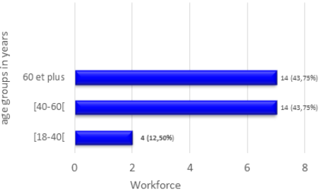
Figure 1.
Distribution of post-stroke aphasic patients by age in years.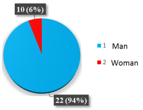
Figure 2.
Distribution of post-stroke aphasic patients by gender.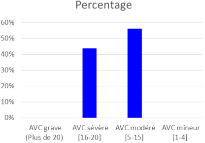
Figure 3.
Distribution of patients according to acute stroke severity.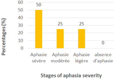
Figure 4.
Distribution of patients according to aphasia severity in the acute phase.
Figure 5.
Pictures to assess oral expression.
Figure 6.
Pictures to assess Listening comprehension.Information

