Introduction: Several reports have been published highlighting accessory root formation in permanent molars. These studies have shown higher incidence of occurrence of accessory roots in patients of Mongolian, Taiwanese, Korean, Inuit, Native American and Indian descent. Very few reports have been published with a focus on accessory root formation in the primary dentition, in particular in the Caucasian population. Objective: This study was aimed at drawing attention to a rare morphological development in the Caucasian population. Case Presentation: This report presents a case of a 13-year-old Caucasian female with a three-rooted mandibular second molar tooth #T. The tooth was surgically removed, with some degree of difficulty, and upon inspection of the tooth following removal, it was observed that an accessory root was present. Conclusions: The presence of an accessory root may be demonstrated radiographically, but often is only discovered after excavation of the pulp chamber or following removal of the tooth itself. As such, it is important for the astute clinician to consider the potential for the presence of an accessory root, and to appreciate the potential complications an accessory may cause during endodontic or surgical treatment. Appropriate imaging should be obtained preoperatively if an accessory root is suspected, and treatment planning should be adjusted accordingly, in order to optimize successful treatment.
This is an Open Access article, distributed under the terms of the Creative Commons Attribution 4.0 International License (http://creativecommons.org/licenses/by/4.0/), which permits unrestricted use, distribution and reproduction in any medium or format, provided the original work is properly cited.
Several reports have been published highlighting accessory root formation in permanent molars. These studies have shown higher incidence of occurrence in patients of Mongolian descent
[1]
Ferraz, J. A. B. J. E. P. Three-rooted mandibular molars in patients of Mongolian, Caucasian and Negro origin. Braz J. 3, 113-117 (1992).
[1]
, specifically Taiwanese
[2]
Tu, M.-G. D., Huang, Heng-Li; et al. Detection of Permanent Three-rooted Mandibular First Molars by Cone-Beam Computed Tomography Imaging in Taiwanese Individuals. JOE 35 (2009).
[2]
, Korean
[3]
Song, J. S., Choi, H. J., Jung, I. Y., Jung, H. S. & Kim, S. O. The prevalence and morphologic classification of distolingual roots in the mandibular molars in a Korean population. J Endod 36, 653-657,
De Moor, R. J. G. The radix entomolaris in mandibular first molars: an endodontic challenge. International endodontic journal 37, 789-799 (2004).
[4]
, Native American
[5]
Turner, C. G. Three rooted mandibular first permanent molars and the question of American Indian Origins. American Journal of Physical Anthropology 34, 229-241 (1971).
[5]
and Indian
[6]
Srivathsa, S. H. Prevalence of three rooted deciduous mandibular molars in Indian children. International Journal of Dental Science and Research 2, 14-16,
. One report published in 1982 demonstrated a three-rooted three-rooted mandibular first molar extracted from a 5 year old Caucasian male, with the accessory root noted to have been located distolingually
[7]
Badger, G. R. Three-rooted mandibular first primary molar. Oral surgery, oral medicine, oral pathology 53, 547 (1982).
[7]
. Given this report, very few reports have been published with a focus on deciduous dentition and fewer present a case of accessory root formation in a Caucasian patient.
The presence of an accessory root may be demonstrated radiographically but often it is only discovered after excavation of the pulp chamber or during and after extraction. As such, it is important for the astute clinician to consider the potential for the presence an accessory root, and to appreciate the potential complications an accessory root may cause during endodontic or surgical intervention. Nevertheless, the implementation of enhanced imaging methods, including cone beam computed tomography (CBCT) has enabled a high degree of sensitivity and specificity in the evolution of surgical and endodontic therapy
[8]
Rios-Osorio, N. et al. Cone-beam computed tomography in endodontics: from the specific technical considerations of acquisition parameters and interpretation to advanced clinical applications. Rstorative Dentistry & Endodontics 49(1): (2024).
This report presents a case of a 13 year old Caucasian female with a three rooted deciduous mandibular second molar tooth T. This purpose of this case report is to draw attention to a rare morphological development in the Caucasian population.
2. Case Report
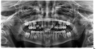
A 13 year old Caucasian female presented for removal of retained tooth T, having been referred for this procedure by her orthodontist. Clinical examination demonstrated healthy 13 year old female, with a mixed dentition in stable condition. Tooth T was noted to be present, with no clinical or radiographic evidence suggestive of accessory root formation (Figure 1).
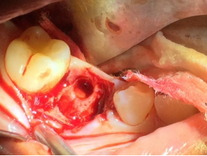
Figure 2. Extraction socket from tooth T, confirming position of accessory root in the distolingual position.
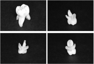
Subsequently, tooth T was removed as treatment planned, in atraumatic fashion, but with some degree of difficulty, in particular due to the expected accentuated curvature typically present in the primary molar dentition. As such, appropriate mucoperiosteal flap development, combined with the judicious use of dental elevators and extraction forceps was employed, in order to minimize the possibility of root or alveolar bone fracture. Upon removal of the tooth, it was clearly apparent that the tooth demonstrated an accessory distolingual root. Inspection of the extraction socket was performed, and confirmed the presence of the accessory root having been located in a distal lingual position (Figure 2). Photographs of the extracted tooth demonstrate the unique nature of the morphology of this tooth, in particular with respect to the accessory distolingual root (Figure 3).
Figure 3. Tooth T viewed from differing aspects following extraction, with accessory distolingual root.
3. Discussion
Deciduous mandibular second molars (K and T) typically present with two roots, one located mesially and one distally. These teeth often closely resemble the permanent mandibular first molars in crown and root morphology
[10]
Nelson, S. J. Wheeler's dental anatomy, physiology and occlusion.. (Elsevier Health Sciences, 2014).
[10]
. Accessory roots form similarly to other roots, by an apical proliferation of the epithelial root sheath
[11]
Thomas, H. F. Root Formation. International Journal of Developmental Biology 39, 231-237 (2003).
[11]
.
The formation of an accessory root is typically considered a variation on the normal tooth anatomy but is not considered pathological
[12]
Song, J. S. et al. Incidence and relationship of an additional root in the mandibular first permanent molar and primary molars. Oral surgery, oral medicine, oral pathology, oral radiology, and endodontics 107, 56-50.
. Goldberg discusses in detail the genetic abnormalities that may contribute to defective root structures, noting that alteration of a chloride channel and/or defective plasminogen gene are linked to local alterations in root formation
[13]
Goldberg, M. Dental Roots: Formation, Lengthening and Malformations of Roots. SOJ Dental and Oral Disorder 1, 1-10 (2021).
. As aforementioned, accessory roots have been reported in greater frequency in those of Mongolian decent. Tu et al analyzed mandibular first molars using CBCT imaging and found that of the 123 patients screened 33% demonstrated an accessory root
[2]
Tu, M.-G. D., Huang, Heng-Li; et al. Detection of Permanent Three-rooted Mandibular First Molars by Cone-Beam Computed Tomography Imaging in Taiwanese Individuals. JOE 35 (2009).
[2]
. Additionally, in a well written literature review by Ahmed et al it was determined that the frequency of maxillary molars with four roots was found to be 0.9%, 1.4% and up to 7% in first, second and third molars respectively.
The research published relative to accessory root formation in primary teeth is more limited. Song et al studied a Korean population and found that out of 4,871 primary mandibular first molars 471 of these (9.7%) demonstrated accessory root formation. Of 5,210 primary mandibular second molars 1,450 (27.8%) demonstrated accessory roots. In a case report published by Rana et al, a 10 year old male Indian patient presented for an extraction of tooth T, it was determined through intraoral periapical radiography and later extraoral clinical examination that an additional root was present in the mesial lingual area
[14]
Rana, V. S. et al. Deciduous mandibular second molar with supernumerary roots and root canals associated with missing mandibular permanent molar. International Journal of Clinical Pediatric Dentistry 4, 167-169 (2011).
[14]
. Very few reports have been published focusing on accessory roots in deciduous teeth of Caucasian children. As noted in the introduction, Badger demonstrated a three-rooted mandibular first molar extracted from a 5 year old Caucasian male. The additional root was shown to be located in the distolingual(DL) area, and he further discussed the fact that the accessory DL root has a predilection towards the right side, and that the existence of a DL root was associated with larger coronal buccolingual dimensions
[7]
Badger, G. R. Three-rooted mandibular first primary molar. Oral surgery, oral medicine, oral pathology 53, 547 (1982).
[7]
. Additionally with respect to associated coronal abnormalities, Andytan et al present presented a case of an accessory cusp being present on bilateral mandibular deciduous first molars in a 6-uear-old male
[15]
Andytan, N. I. I., et al. An Accessory Cusp and Three-rooted Deciduous Mandibular First Molar: A Rare Entity. Journal of Pediatric Dentistry 9, 32-26 (2023).
. With respect to the rarity of an accessory root in the in the Caucasian population, this would appear to be the result of genetic factors, with possible epigenetic influences, and presents a unique opportunity for further research.
4. Conclusion
The present case report discusses a deciduous second molar (tooth T) with an accessory root, discovered upon extraction. This is significant because it confirms the possibility of accessory primary roots in the Caucasian population. The occurrence of such accessory roots can impact treatment in endodontic therapy and surgical removal. In particular, the canal of an accessory root canal may be missed during endodontic treatment, or a portion of a fractured root may be left in situ, both of which might result in adverse sequelae. As such, it is strongly recommended that if accessory root(s) are suspected upon presentation, additional imaging modalities should be considered, including cone beam computed tomography (CBCT) scanning and periapical radiography, in order to aid in diagnosis and treatment planning. Additionally, the presence of an unrecognized accessory root can significantly increase the difficulty of endodontic therapy, including pulpotomy/pulpectomy, or surgical removal. The astute clinician should keep in mind the potential accessory root formation, even in the face of clinical and radiographic findings that may not suggest the presence of accessory root formation.
Abbreviations
CBCT
Cone Beam Computed Tomography
DL
Distolingual
Author Contributions
Daniel Joseph Traub: Funding acquisition, Resources, Software
Robert Walsh: Conceptualization, Data curation, Formal Analysis, Funding acquisition, Investigation, Project administration, Resources, Software, Supervision, Validation, Visualization
Ferraz, J. A. B. J. E. P. Three-rooted mandibular molars in patients of Mongolian, Caucasian and Negro origin. Braz J. 3, 113-117 (1992).
[2]
Tu, M.-G. D., Huang, Heng-Li; et al. Detection of Permanent Three-rooted Mandibular First Molars by Cone-Beam Computed Tomography Imaging in Taiwanese Individuals. JOE 35 (2009).
[3]
Song, J. S., Choi, H. J., Jung, I. Y., Jung, H. S. & Kim, S. O. The prevalence and morphologic classification of distolingual roots in the mandibular molars in a Korean population. J Endod 36, 653-657,
De Moor, R. J. G. The radix entomolaris in mandibular first molars: an endodontic challenge. International endodontic journal 37, 789-799 (2004).
[5]
Turner, C. G. Three rooted mandibular first permanent molars and the question of American Indian Origins. American Journal of Physical Anthropology 34, 229-241 (1971).
[6]
Srivathsa, S. H. Prevalence of three rooted deciduous mandibular molars in Indian children. International Journal of Dental Science and Research 2, 14-16,
Badger, G. R. Three-rooted mandibular first primary molar. Oral surgery, oral medicine, oral pathology 53, 547 (1982).
[8]
Rios-Osorio, N. et al. Cone-beam computed tomography in endodontics: from the specific technical considerations of acquisition parameters and interpretation to advanced clinical applications. Rstorative Dentistry & Endodontics 49(1): (2024).
Nelson, S. J. Wheeler's dental anatomy, physiology and occlusion.. (Elsevier Health Sciences, 2014).
[11]
Thomas, H. F. Root Formation. International Journal of Developmental Biology 39, 231-237 (2003).
[12]
Song, J. S. et al. Incidence and relationship of an additional root in the mandibular first permanent molar and primary molars. Oral surgery, oral medicine, oral pathology, oral radiology, and endodontics 107, 56-50.
Rana, V. S. et al. Deciduous mandibular second molar with supernumerary roots and root canals associated with missing mandibular permanent molar. International Journal of Clinical Pediatric Dentistry 4, 167-169 (2011).
[15]
Andytan, N. I. I., et al. An Accessory Cusp and Three-rooted Deciduous Mandibular First Molar: A Rare Entity. Journal of Pediatric Dentistry 9, 32-26 (2023).
Traub, D. J., Walsh, R., Ahern, C. (2025). A Three-rooted Deciduous Second Molar in a 13-year-old Caucasian Female. International Journal of Medical Case Reports, 4(3), 51-54. https://doi.org/10.11648/j.ijmcr.20250403.13
Traub, D. J.; Walsh, R.; Ahern, C. A Three-rooted Deciduous Second Molar in a 13-year-old Caucasian Female. Int. J. Med. Case Rep.2025, 4(3), 51-54. doi: 10.11648/j.ijmcr.20250403.13
Traub DJ, Walsh R, Ahern C. A Three-rooted Deciduous Second Molar in a 13-year-old Caucasian Female. Int J Med Case Rep. 2025;4(3):51-54. doi: 10.11648/j.ijmcr.20250403.13
@article{10.11648/j.ijmcr.20250403.13,
  author = {Daniel Joseph Traub and Robert Walsh and Colleen Ahern},
  title = {A Three-rooted Deciduous Second Molar in a 13-year-old Caucasian Female
},
  journal = {International Journal of Medical Case Reports},
  volume = {4},
  number = {3},
  pages = {51-54},
  doi = {10.11648/j.ijmcr.20250403.13},
  url = {https://doi.org/10.11648/j.ijmcr.20250403.13},
  eprint = {https://article.sciencepublishinggroup.com/pdf/10.11648.j.ijmcr.20250403.13},
  abstract = {Introduction: Several reports have been published highlighting accessory root formation in permanent molars. These studies have shown higher incidence of occurrence of accessory roots in patients of Mongolian, Taiwanese, Korean, Inuit, Native American and Indian descent. Very few reports have been published with a focus on accessory root formation in the primary dentition, in particular in the Caucasian population. Objective: This study was aimed at drawing attention to a rare morphological development in the Caucasian population. Case Presentation: This report presents a case of a 13-year-old Caucasian female with a three-rooted mandibular second molar tooth #T. The tooth was surgically removed, with some degree of difficulty, and upon inspection of the tooth following removal, it was observed that an accessory root was present. Conclusions: The presence of an accessory root may be demonstrated radiographically, but often is only discovered after excavation of the pulp chamber or following removal of the tooth itself. As such, it is important for the astute clinician to consider the potential for the presence of an accessory root, and to appreciate the potential complications an accessory may cause during endodontic or surgical treatment. Appropriate imaging should be obtained preoperatively if an accessory root is suspected, and treatment planning should be adjusted accordingly, in order to optimize successful treatment.},
 year = {2025}
}
TY  - JOUR
T1  - A Three-rooted Deciduous Second Molar in a 13-year-old Caucasian Female

AU  - Daniel Joseph Traub
AU  - Robert Walsh
AU  - Colleen Ahern
Y1  - 2025/08/20
PY  - 2025
N1  - https://doi.org/10.11648/j.ijmcr.20250403.13
DO  - 10.11648/j.ijmcr.20250403.13
T2  - International Journal of Medical Case Reports
JF  - International Journal of Medical Case Reports
JO  - International Journal of Medical Case Reports
SP  - 51
EP  - 54
PB  - Science Publishing Group
SN  - 2994-7049
UR  - https://doi.org/10.11648/j.ijmcr.20250403.13
AB  - Introduction: Several reports have been published highlighting accessory root formation in permanent molars. These studies have shown higher incidence of occurrence of accessory roots in patients of Mongolian, Taiwanese, Korean, Inuit, Native American and Indian descent. Very few reports have been published with a focus on accessory root formation in the primary dentition, in particular in the Caucasian population. Objective: This study was aimed at drawing attention to a rare morphological development in the Caucasian population. Case Presentation: This report presents a case of a 13-year-old Caucasian female with a three-rooted mandibular second molar tooth #T. The tooth was surgically removed, with some degree of difficulty, and upon inspection of the tooth following removal, it was observed that an accessory root was present. Conclusions: The presence of an accessory root may be demonstrated radiographically, but often is only discovered after excavation of the pulp chamber or following removal of the tooth itself. As such, it is important for the astute clinician to consider the potential for the presence of an accessory root, and to appreciate the potential complications an accessory may cause during endodontic or surgical treatment. Appropriate imaging should be obtained preoperatively if an accessory root is suspected, and treatment planning should be adjusted accordingly, in order to optimize successful treatment.
VL  - 4
IS  - 3
ER  -